AI in Medical Imaging: How It Works, Applications, Benefits, and Challenges
Artificial intelligence has rapidly changed the world we live in, creating new possibilities across industries and reshaping how many everyday and professional tasks are performed. One of the most promising areas of this transformation is healthcare, where the use of AI in medical imaging is giving doctors and other healthcare professionals new ways to analyze complex imaging data, automate certain tasks, and support clinical decision-making.
But what does AI actually bring to medical imaging, and where are its limits? In this article, we explore how artificial intelligence is used in medical imaging, its major applications, benefits, and challenges, and answer an important question surrounding its growing role in healthcare: Could AI eventually replace radiologists and other medical imaging professionals?
What Is AI in Medical Imaging?
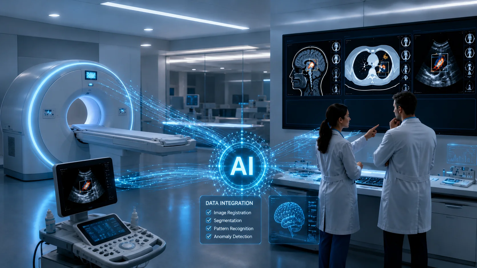
AI in medical imaging refers to the use of artificial intelligence to analyze, process, and extract useful information from medical images. AI is a broad field that includes technologies such as machine learning and deep learning, which empower computer systems to learn patterns from data and perform specific tasks. In medical imaging, these technologies can analyze X-rays, CT scans, MRI, mammography, ultrasound, PET, and other imaging data to support tasks such as detection, segmentation, measurement, image processing, and quantitative analysis. As medical imaging generates increasingly large and complex datasets, AI can help healthcare professionals handle analytical and repetitive tasks more efficiently and consistently.
However, the role of AI in medical imaging extends beyond analyzing images after they are generated. AI can support various phases of the imaging workflow, including image acquisition, reconstruction, processing, and even transducer quality assurance, where technologies such as a smart transducer analyzer can help evaluate ultrasound transducer performance.
As these AI technologies become more integrated into healthcare environments, their value depends not only on how well an algorithm performs but also on how effectively it fits into clinical workflows and supports professional judgment. To understand exactly how AI works in medical imaging, continue to the next section, where we break down the process and the technologies behind it.
How AI Works in Medical Imaging
AI in medical imaging uses algorithms trained on imaging data to recognize patterns and extract relevant information from medical images. These algorithms can be integrated into the software of imaging systems, including certain new and refurbished ultrasound machines, depending on the model, software version, and configuration.
Two key technologies behind many of these AI capabilities are machine learning (ML) and its more advanced subset, deep learning (DL).
Machine learning (ML)
Machine learning (ML) is a branch of artificial intelligence (AI) that allows computer systems to learn patterns and relationships from data rather than relying only on explicitly programmed rules. In medical imaging, ML models can be trained using information from medical images to recognize relevant patterns. Once trained, these models can apply what they have learned to new images and perform specific tasks, such as identifying abnormalities, classifying findings, or assisting with image analysis.
Deep learning (DL)
Deep learning is a bit more complicated than machine learning and uses neural networks with multiple layers to learn complex patterns directly from large amounts of data. One important type is the convolutional neural network (CNN), a deep-learning model designed to recognize spatial and visual patterns in images. In medical imaging, CNNs can learn increasingly complex image models and features, from basic shapes and edges to patterns associated with anatomical structures or abnormalities.
Ultrasound is just one area where these technologies are applied. AI is used across various medical imaging modalities, with some of its most important applications discussed in the next section.
Applications of AI Across Medical Imaging Modalities
By now, you should have a clear idea of what AI in medical imaging is and how it works. However, you might wonder what it looks like in practice.
Well, some of the most important use cases of this technology are explained as follows.
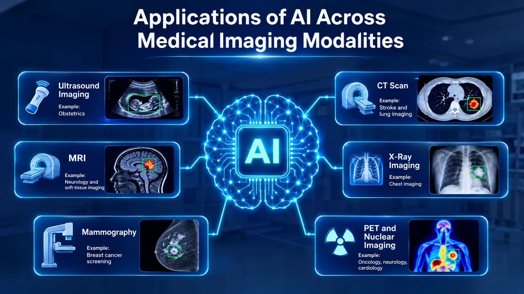
AI in Ultrasound Imaging
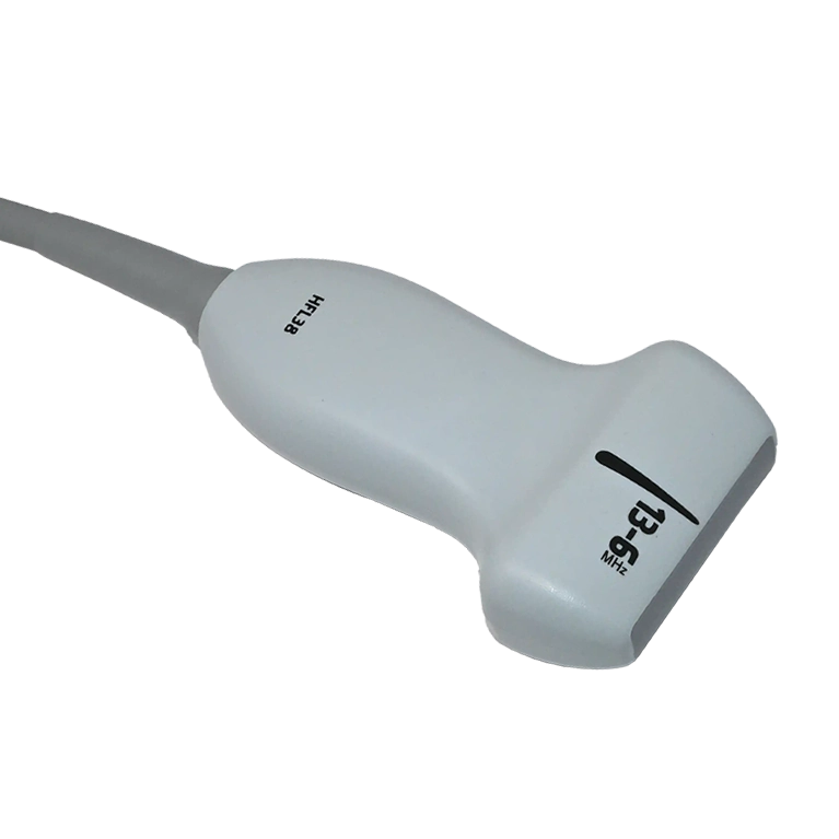
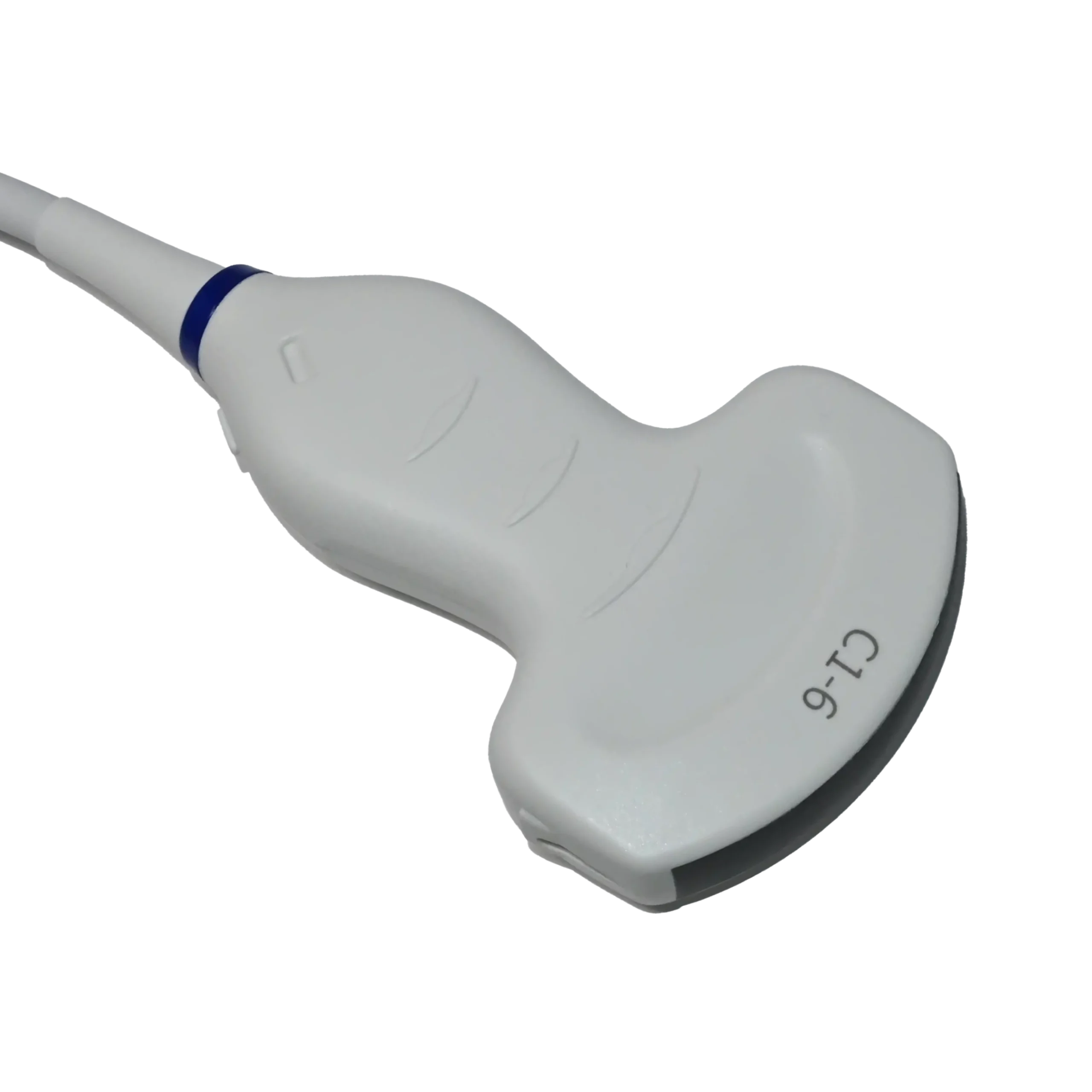
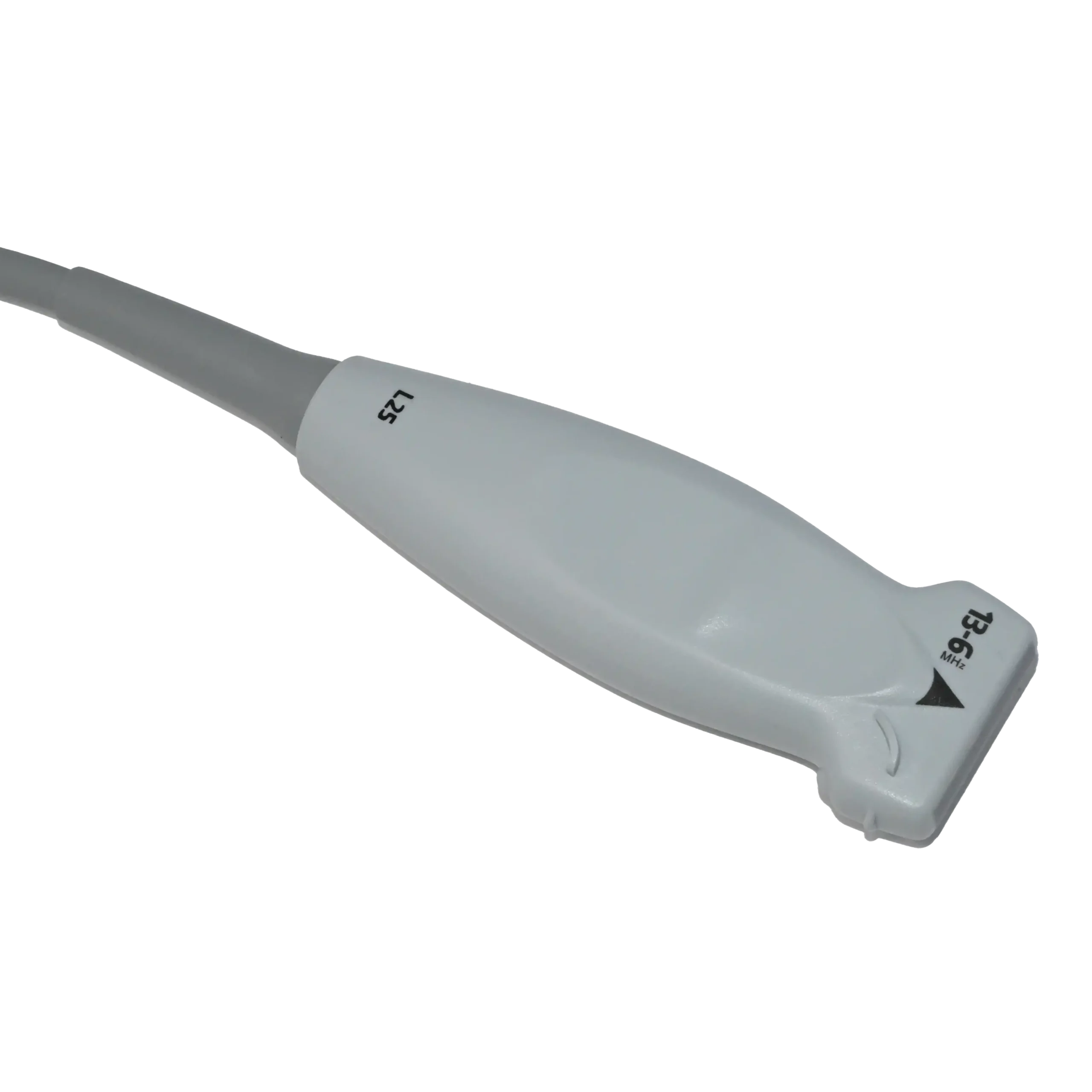
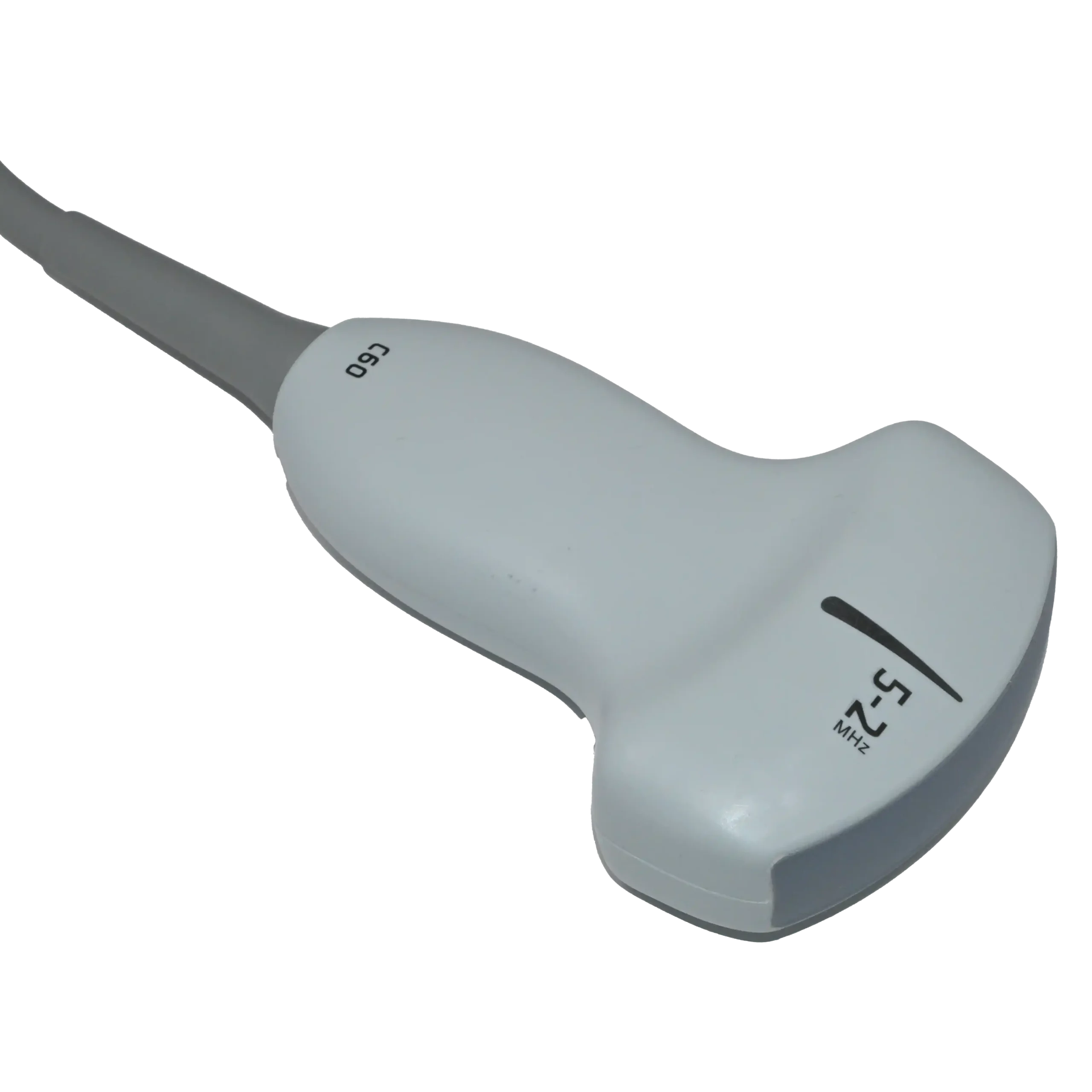
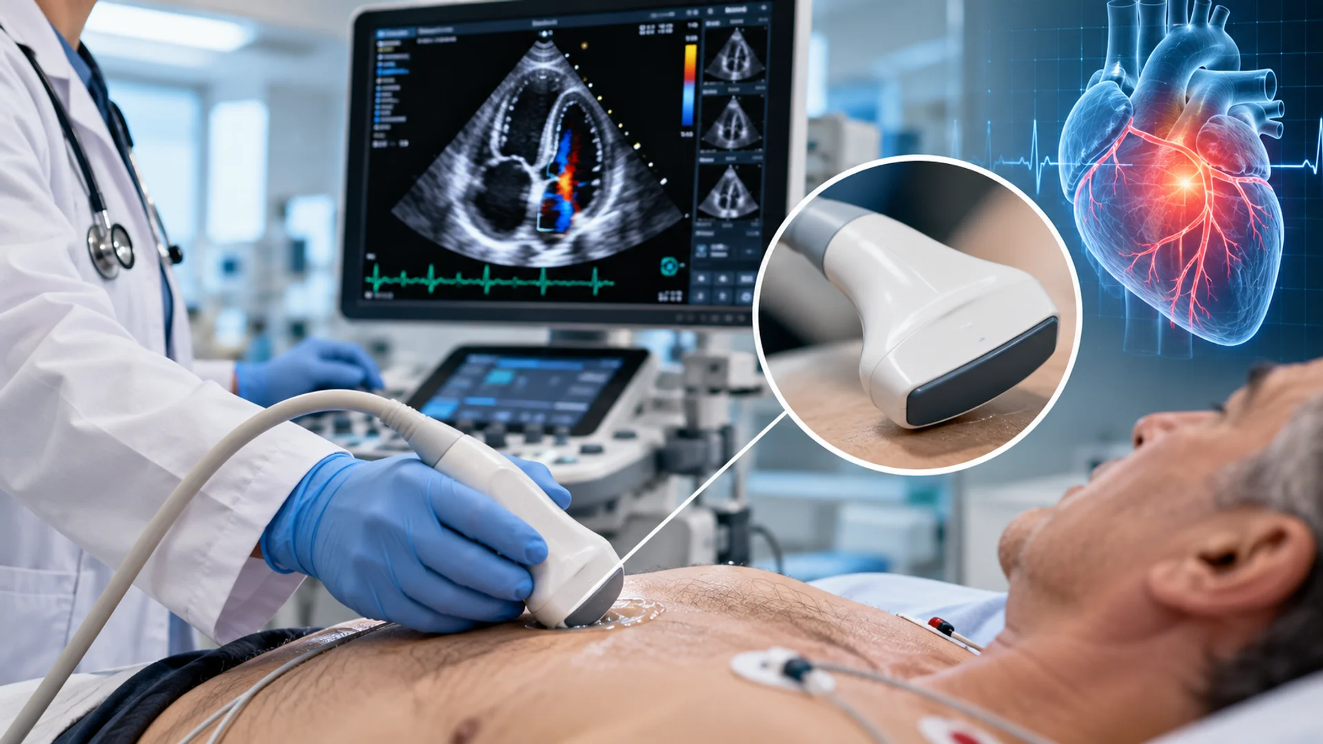
Ultrasound machines use different types of ultrasound transducers to send and receive high-frequency sound waves, producing real-time images of organs, tissues, blood flow, and other structures within the body. AI can work with these images to support various aspects of image acquisition and analysis.
These technologies can assist with recognizing anatomical structures, taking automated measurements, analyzing lesions, supporting image acquisition, and evaluating image quality.
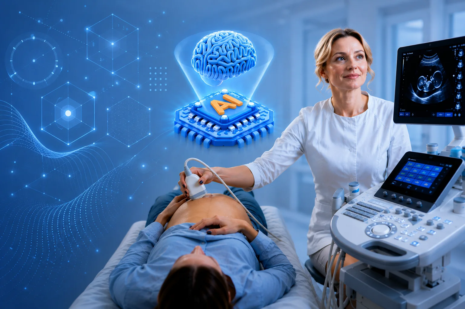
In obstetric ultrasound, for example, AI has been studied for identifying standard imaging views and assisting with fetal measurements. AI may also help assess image quality, supporting more consistent ultrasound examinations.
AI in X-Ray Imaging
In X-ray imaging, doctors use ionizing radiation to create two-dimensional images of structures inside the body. In this field, AI can analyze X-rays to help identify patterns associated with conditions such as fractures, lung nodules, and certain chest abnormalities.
Depending on the system, it may highlight suspicious regions or classify findings that require closer review and examination, giving healthcare professionals additional information when performing an X-Ray image.
AI in CT Imaging
While conventional X-rays produce two-dimensional images, computed tomography (CT) combines X-ray measurements taken from multiple angles to create detailed cross-sectional images of the body.
Regarding CT imaging, AI can help recognize and characterize abnormalities, segment organs and lesions, and deliver automated measurements on CT images. It is also being applied to image reconstruction and noise reduction, expanding its role beyond the detection of abnormalities alone.
AI in MRI
Unlike X-ray and CT, magnetic resonance imaging (MRI) uses magnetic fields and radiofrequency signals rather than ionizing radiation to produce detailed images, particularly of soft tissues. In this field, AI is used to segment anatomical structures and lesions, perform measurements, and detect relevant image patterns. AI-based reconstruction methods are also being investigated to generate useful images from less acquired data, which may help shorten certain MRI examinations.
AI in Mammography
Mammography is a specialized type of X-ray imaging primarily used for breast cancer screening and evaluation. AI can analyze mammograms to identify suspicious regions, assist with lesion detection and classification, and support assessments such as breast density and cancer risk.
AI in PET and Nuclear Imaging
Unlike imaging methods that mainly show anatomical structures, nuclear medicine provides information about how organs and tissues are functioning. It uses small amounts of radioactive substances, known as radiotracers, to show physiological and molecular activity within the body.
Positron emission tomography (PET) is one of the main imaging techniques used in nuclear medicine. AI is increasingly being explored in PET and other nuclear imaging applications to improve image reconstruction, reduce noise, detect and outline lesions, and support quantitative analysis. These capabilities can help healthcare professionals evaluate biological activity and disease processes in areas such as cancer care, neurology, and cardiology.
To sum up, here’s a quick comparison of how AI is being used across these medical imaging modalities:
| Imaging Modality | Example Clinical Area | What AI Can Support |
| Ultrasound | Obstetrics | Recognizing standard views and assisting with fetal measurements |
| X-Ray | Chest imaging | Drawing attention to potentially abnormal findings |
| CT Scan | Stroke and lung imaging | Identifying relevant patterns and quantifying abnormalities |
| MRI | Neurology and soft-tissue imaging | Analyzing complex structures and supporting faster reconstruction |
| Mammography | Breast cancer screening | Identifying areas that may require closer assessment |
| PET | Oncology, neurology, cardiology | Analyzing functional and molecular imaging information |
Benefits of AI in Medical Imaging
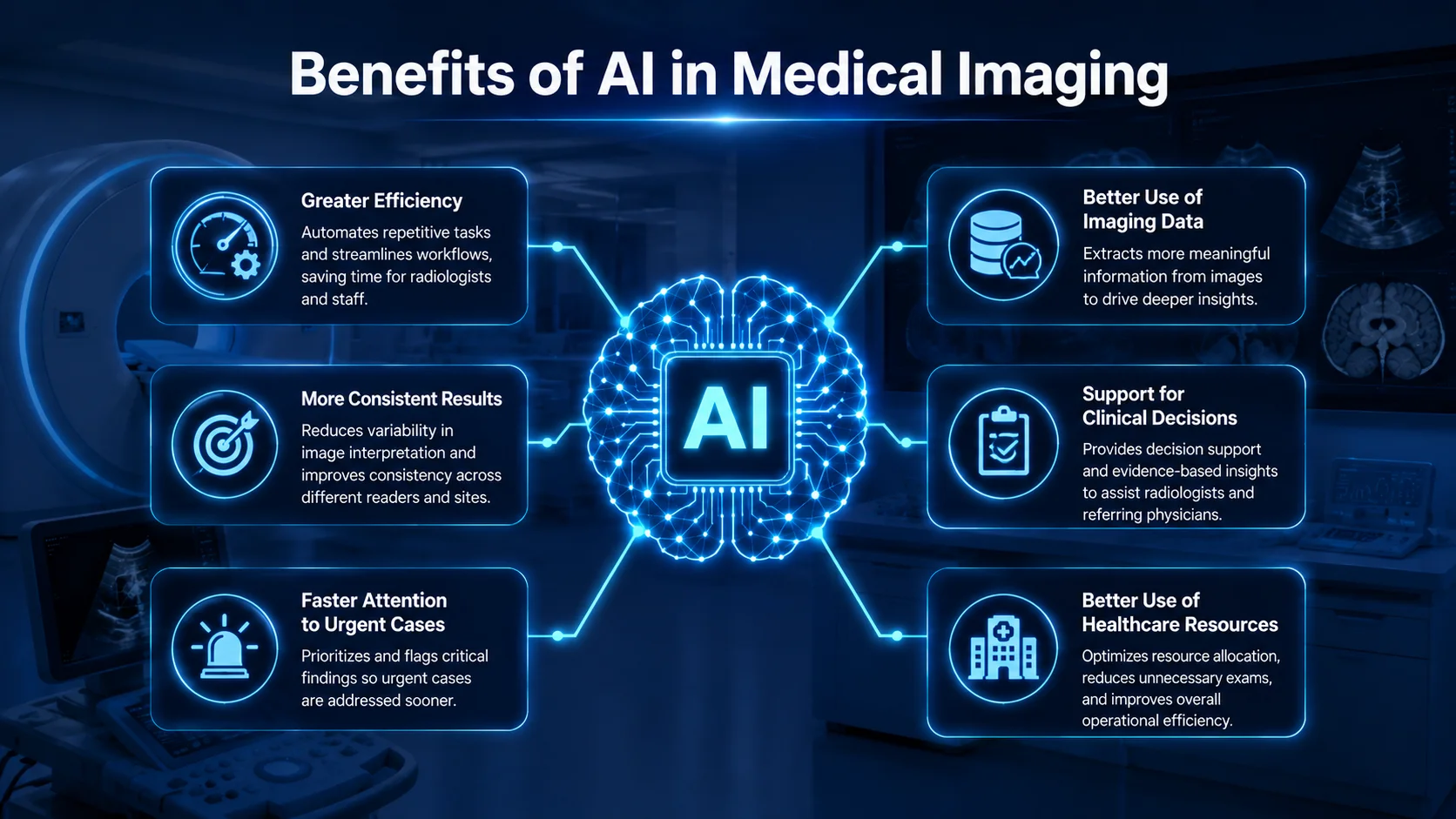
Looking at the medical applications above shows just how broadly AI can be used across medical imaging, from machines using new and refurbished ultrasound transducers to modalities such as X-ray, CT, MRI, mammography, and PET. However, the value of AI goes beyond the imaging process itself, with potential benefits for healthcare professionals, imaging departments, and ultimately patient care.
- Greater Efficiency: AI can reduce the time spent on selected routine tasks and help professionals focus on more complex work.
- More Consistent Results: Applying standardized methods can reduce variation in certain measurements and image assessments.
- Faster Attention to Urgent Cases: AI-supported prioritization may help potentially critical examinations reach clinicians sooner.
- Better Use of Imaging Data: AI can extract quantitative information and patterns that add another layer of information to image evaluation.
- Support for Clinical Decisions: AI-generated insights can provide additional information for clinicians when assessing imaging findings.
- Better Use of Healthcare Resources: Automating suitable tasks can help imaging departments use available expertise and resources more effectively.
Importance of Image and Data Quality for AI in Medical Imaging
The benefits of AI in medical imaging depend on more than the sophistication of an algorithm or its ability to recognize patterns. AI models learn from data and analyze the images they receive, meaning that the quality, consistency, and relevance of these inputs can directly affect how accurately and reliably the models perform.
Image Quality
One important factor when using AI in medical imaging is the quality of the images being analyzed. Noise, artifacts, patient movement, acquisition settings, and equipment can all affect image quality. For example, with an OB/GYN ultrasound machine, probe positioning and patient or fetal movement can influence the images available for AI analysis.
Because AI models rely on visual features within these images, such variations can affect their output. Maintaining consistent image quality is therefore important for both clinical interpretation and the reliable use of AI in medical imaging.
Training Data Quality
The reliability of AI in medical imaging also depends heavily on the data used to develop and train the model. Training data should have accurate labels and include enough relevant examples to help the model learn meaningful patterns.
A large dataset alone is not enough if it does not represent the patients, medical conditions, and imaging settings where the AI will be used. Using accurate and representative data is therefore important for reliable AI performance in clinical practice.
Consistency Across Imaging Systems
Medical images can vary across hospitals and healthcare settings. Differences in scanners, equipment, imaging protocols, acquisition settings, and patient populations can affect the images that an AI system analyzes. Because of these differences, an AI model that performs well in one setting may not perform as reliably in another. Testing the model with data from different clinical settings and maintaining consistent imaging practices are therefore important for its reliable use in real-world healthcare.
Challenges of AI in Medical Imaging
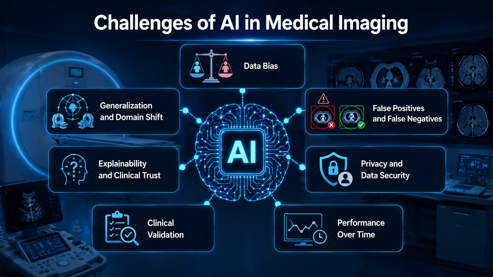
As mentioned earlier, the use of AI in medical imaging comes with many potential benefits, from reducing repetitive tasks to supporting image interpretation and time-sensitive care. However, these benefits do not come automatically. The performance of an AI system depends on the data it learns from, the environment in which it is used, and how effectively it is validated and integrated into clinical practice. Several challenges therefore need to be considered alongside its potential advantages.
- Data Bias: AI models may be less reliable when used with patients or clinical conditions that are underrepresented in their training data.
- Generalization and Domain Shift: Differences in scanners, imaging protocols, and clinical environments can affect AI models’ performance. A model that works well in one setting may not perform equally well in another.
- False Positives and False Negatives: AI can incorrectly flag a normal finding or miss an abnormal one. Its results therefore need appropriate clinical review.
- Explainability and Clinical Trust: Some AI models can deliver results without clearly showing how they reached their conclusions. This lack of transparency can make it challenging for clinicians to understand the reasoning behind an AI-generated result and consider how much confidence to place in it.
- Privacy and Data Security: Medical images can involve sensitive patient information. Strong safeguards are therefore necessary when imaging data is stored, shared, or processed by AI model systems in medical imaging.
- Clinical Validation: Good performance during AI development does not always mean that the model will perform equally well in real clinical settings. For this reason, AI models should be tested using independent data and evaluated in the healthcare settings where they are intended to be used.
- Performance Over Time: Changes in ultrasound equipment, software, imaging protocols, or patient populations can affect how an AI system performs over time. Regular monitoring is therefore important to ensure that the system continues to work reliably and as expected in clinical practice.
The Future of AI in Medical Imaging: Will AI Replace Radiologists?
As AI becomes more capable, some may wonder whether it could eventually replace radiologists or other healthcare professionals. However, medical imaging involves much more than analyzing images; it requires clinical expertise to understand what those images mean in the context of each patient. This is true even in routine examinations such as an abdominal ultrasound imaging, where imaging findings must be evaluated alongside the patient’s symptoms, medical history, and other clinical information.
For this reason, AI is more likely to work alongside healthcare professionals than replace them. It can assist with image analysis, measurements, and pattern recognition, but radiologists, physicians, and technologists provide the clinical knowledge and professional judgment needed to interpret results in context. AI is therefore better viewed as a tool that supports medical expertise rather than replaces it.
Conclusion
AI in medical imaging is expanding how healthcare professionals analyze images, manage imaging data, and support clinical decisions. From image analysis and automated measurements to workflow support, its potential continues to grow, but reliable performance still depends on high-quality data, proper validation, and appropriate human oversight.
Rather than replacing medical imaging professionals, AI is becoming a tool that can complement their expertise. As the technology continues to advance, combining AI capabilities with professional judgment and a strong focus on patient safety will remain essential.