Breast ultrasound vs mammogram: which one is better?
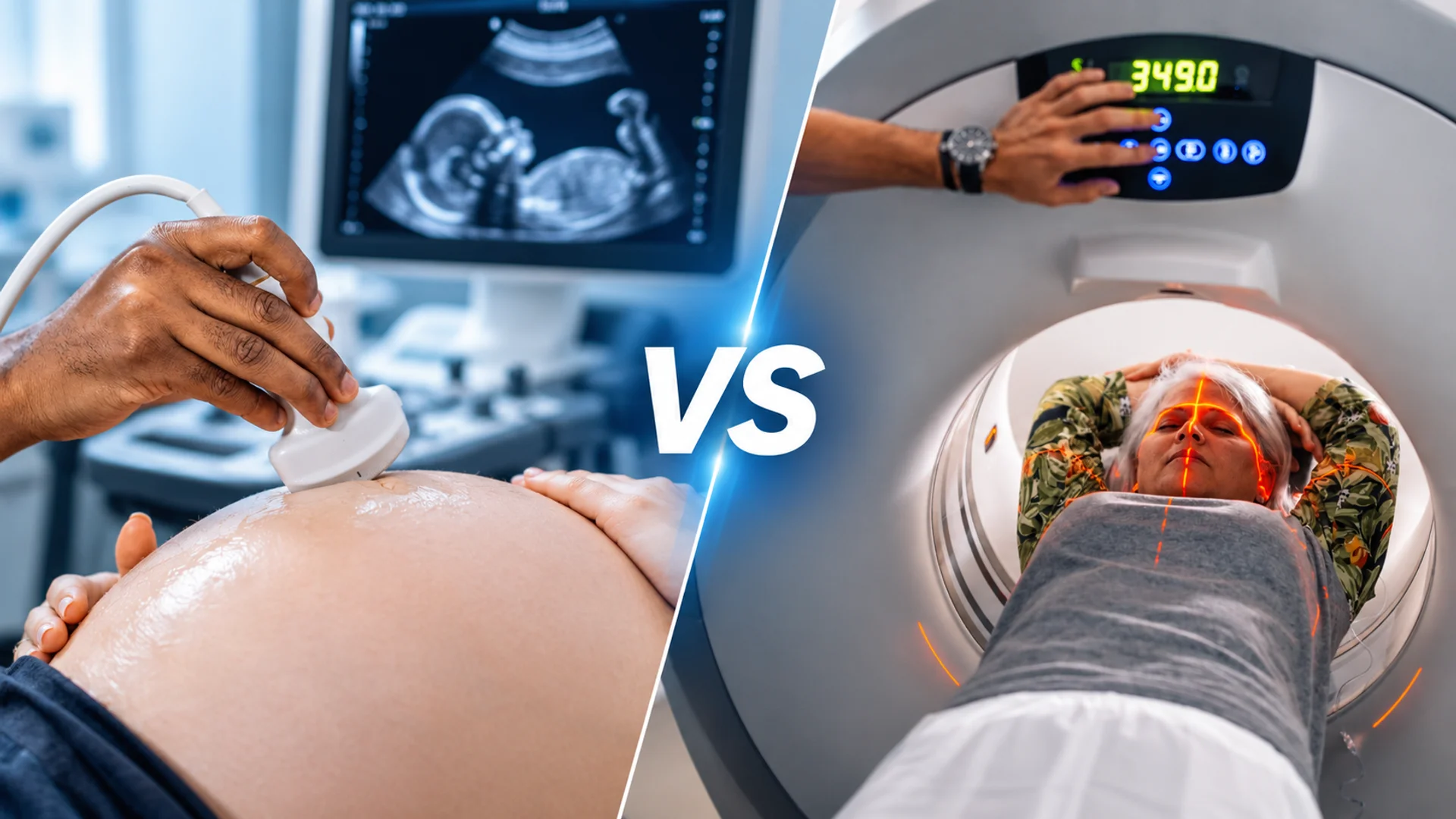
Although some people may think breast ultrasound and mammography are the same examination, they are two distinct imaging methods with different clinical applications.
Breast ultrasound is commonly best for identifying breast lumps and abnormalities, while mammography is primarily used for routine breast cancer screening and can reveal findings such as microcalcifications.
This guide compares mammograms and breast ultrasound, explaining how each works, what it can detect, and when doctors may recommend one or both.
Breast Ultrasound vs Mammogram Quick Comparison
Breast ultrasound and mammography are two commonly used medical imaging methods for evaluating breast health, but each has different capabilities and clinical applications. Here is a quick overview of how they compare.
| Comparison factor | Breast Ultrasound | Mammogram |
| Imaging technology | Uses high-frequency sound waves | Uses low-dose X-rays |
| Image production | Produces real-time images | Produces 2D images or 3D reconstructions |
| Main purpose | Diagnostic evaluation and supplemental screening | Routine breast cancer screening and diagnostic evaluation |
| Radiation exposure | No ionizing radiation | Uses ionizing radiation |
| Breast compression | No compression | Requires breast compression |
| Examination method | Handheld transducer moves over the breast | Breast is positioned between compression plates |
| Examination duration | Usually takes around 15–30 minutes | Usually takes around 15–30 minutes |
| Breast lumps | Helps examine lump composition and internal structure | Shows the lump’s location and surrounding tissue |
| Breast cysts | Particularly effective at identifying fluid-filled cysts | Particularly effective at identifying and assessing calcification patterns |
| Microcalcifications | Less reliable for detecting tiny calcium deposits | Particularly effective at identifying and assessing calcification patterns |
| Dense breast tissue | Can reveal additional abnormalities hidden by dense tissue | Dense tissue can obscure some abnormalities |
| Breast cancer screening | Not a standalone replacement for routine mammography | Primary screening method for most women at average risk |
| Breast implants | Useful for evaluating certain implant complications | Used for cancer screening with special implant-displacement views |
| Pregnancy | Used for cancer screening with special implant-displacement views | Can be performed when clinically indicated |
| Cost | Often less expensive, depending on location and provider | May cost more, especially for diagnostic or 3D examinations |
Breast Ultrasound vs. Mammogram: How Does Each Work?
To compare breast ultrasound vs mammogram, let’s first look at how each imaging method works. Understanding how ultrasound machines and mammogram systems work will make their key differences easier to understand.
How Does a Breast Ultrasound Work?
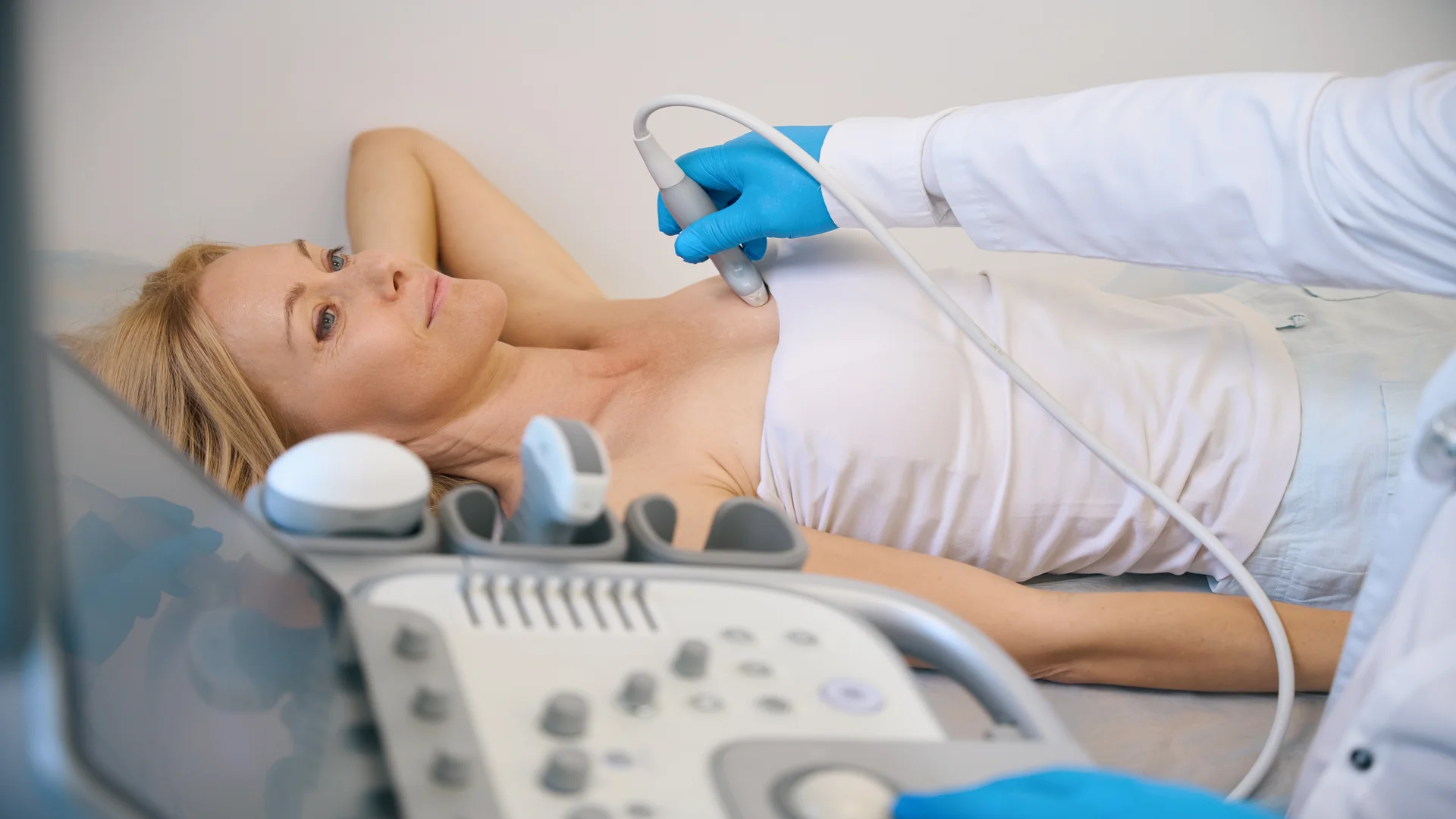
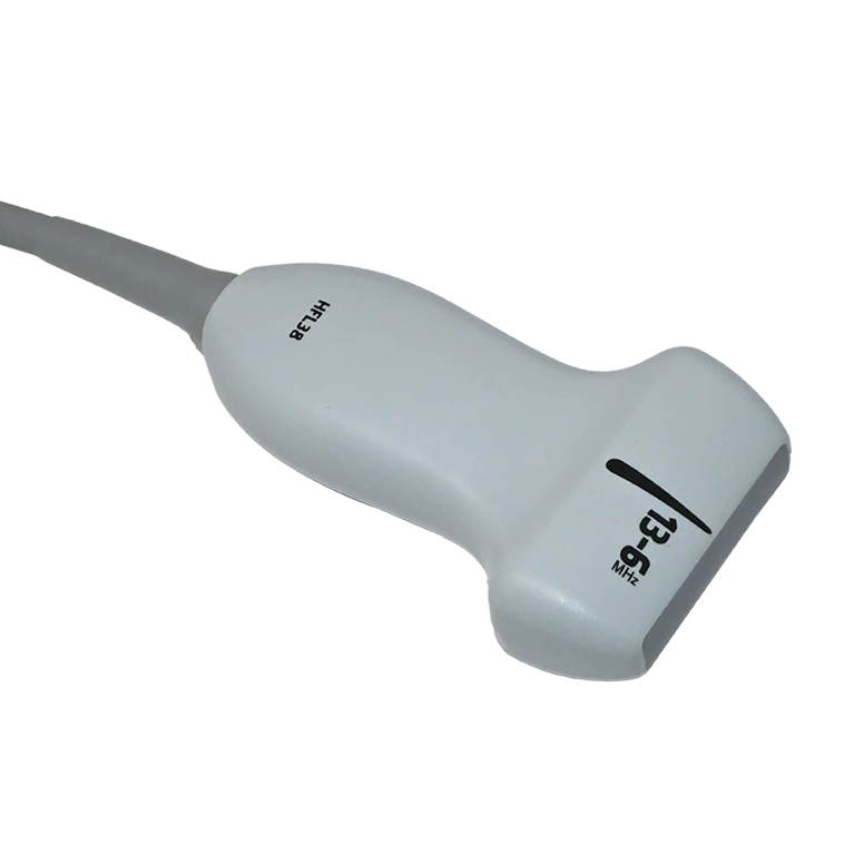
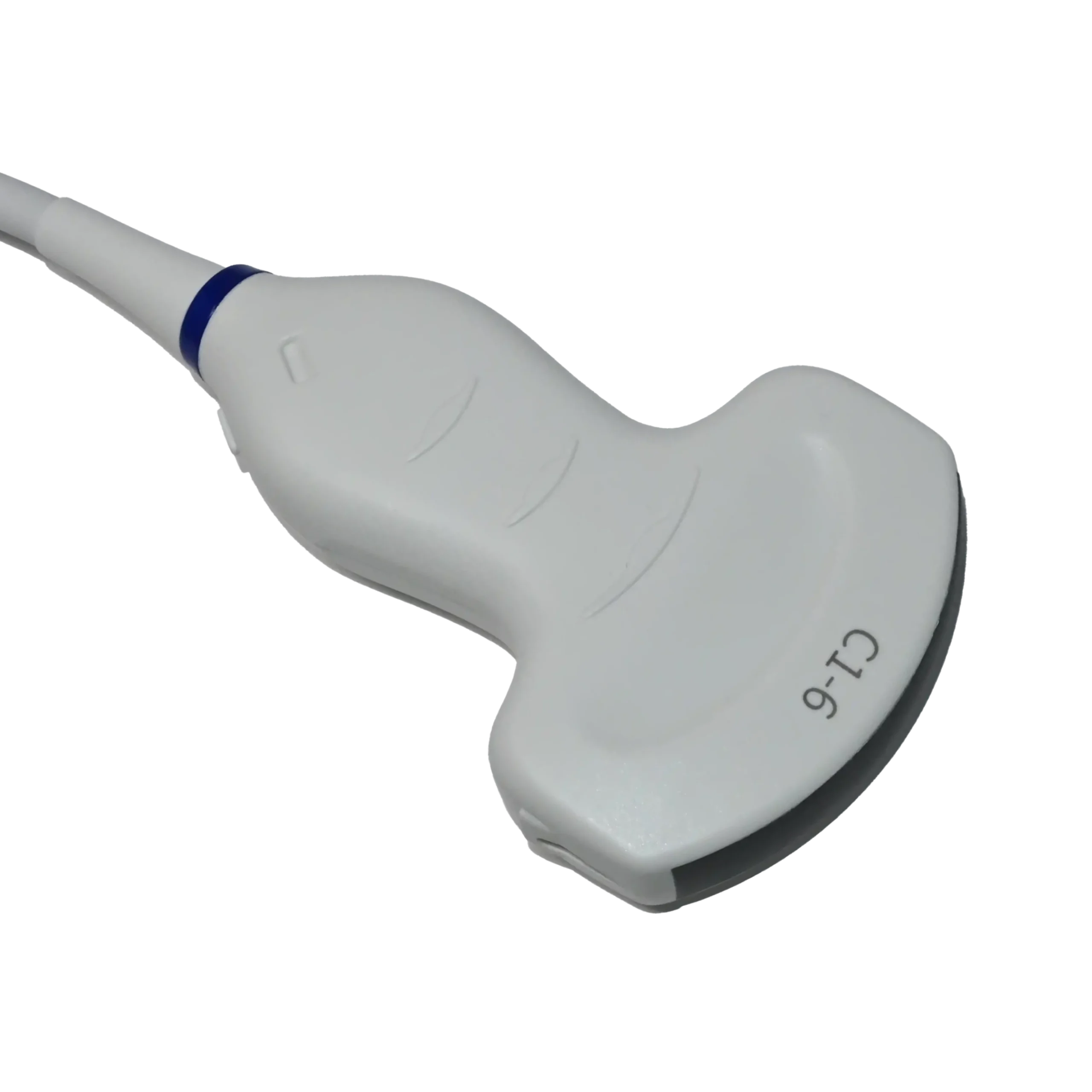
As the name suggests, this examination uses an ultrasound machine and a suitable transducer.
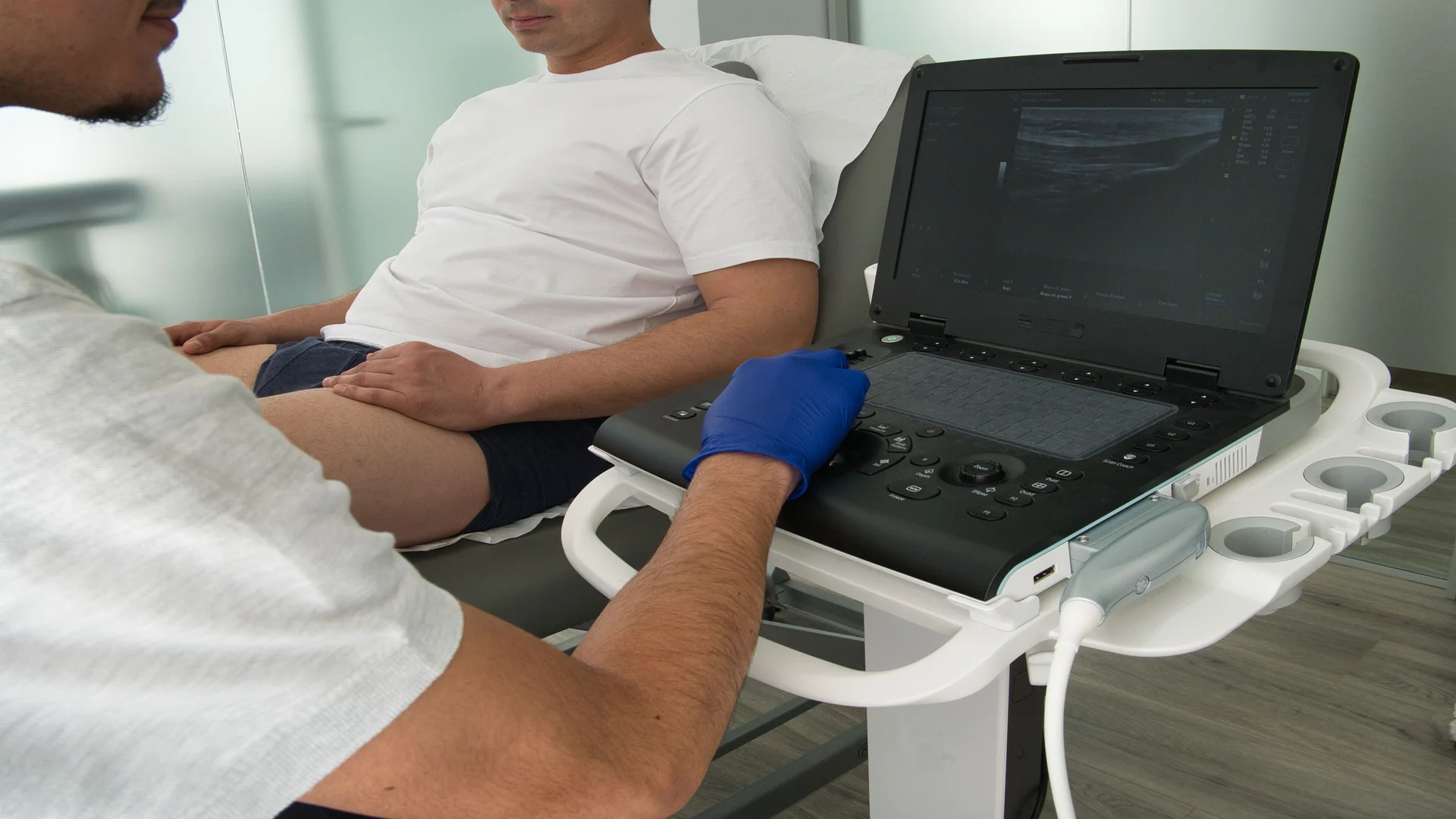
During a breast examination, a sonographer applies gel to the skin and moves a breast ultrasound probe over the breast. The probe sends high-frequency sound waves into the tissue and receives the returning echoes. The ultrasound machine then processes these echoes and displays them as real-time images. Depending on the clinical setting, the examination can be performed using either portable or console-based ultrasound machines.
Alternatively, automated whole-breast ultrasound (ABUS) uses a specialized scanning system to capture images across a larger area of the breast.
How Does a Mammogram Work?
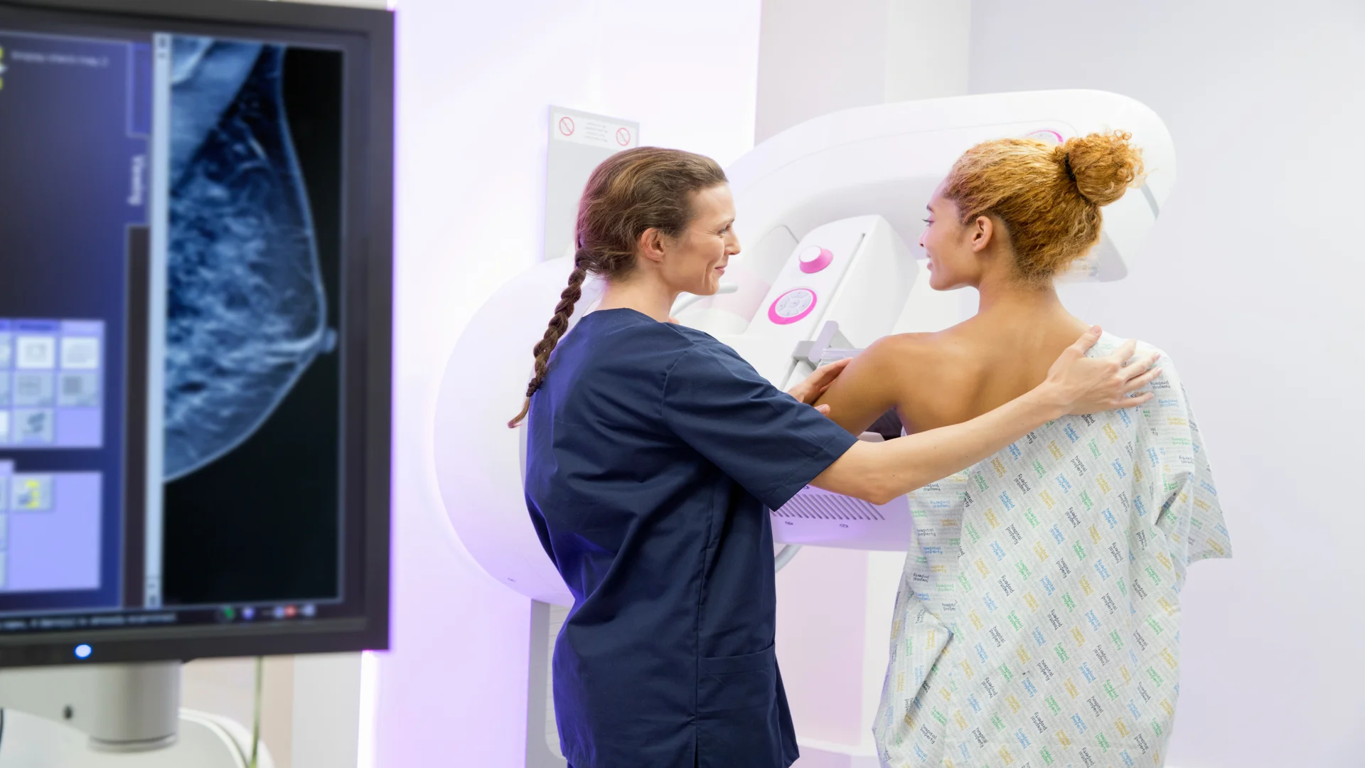
A mammogram uses low-dose X-rays to create images of breast tissue. During the examination, each breast is placed on an imaging platform and compressed between two plates to spread the tissue and improve image clarity.
Standard screening typically captures two views of each breast. While 2D mammography produces flat images, 3D mammography, also called digital breast tomosynthesis, captures multiple images from different angles to create thin slices of breast tissue.
Breast Ultrasound vs. Mammogram: 7 Key Differences
Now that you understand how both imaging methods work, let’s compare breast ultrasound vs. mammogram from key aspects. Although both examine breast tissue, their technologies, image acquisition methods, and clinical applications differ.
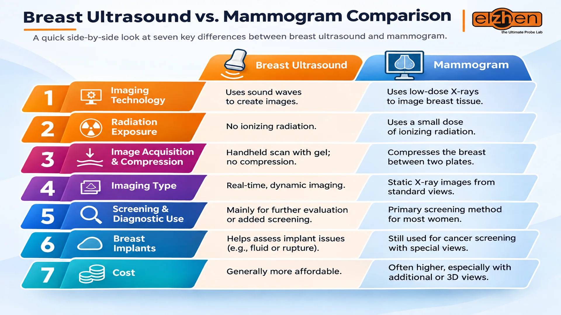
1. Imaging Technology
The main difference between these examinations is the technology they use to produce images.
- Breast ultrasound: Uses high-frequency sound waves transmitted through breast tissue by a transducer. A computer processes the returning echoes to create images.
- Mammogram: Uses low-dose X-rays to capture images of the internal structure of the breast.
2. Radiation Exposure
Another important factor to consider when comparing the mammogram vs breast ultrasound is whether the examination involves ionizing radiation.
- Breast ultrasound: Does not use ionizing radiation. As a result, it’s a better choice for repeated diagnostic examinations when clinically needed.
- Mammogram: Uses a low dose of ionizing radiation to produce breast images. The benefits of this medical scan generally outweigh the small potential risk from radiation exposure.
3. Image Acquisition and Breast Compression
The way images are captured also differs, particularly in how the breast is positioned during each examination.
- Breast ultrasound: The sonographer moves a handheld transducer over the breast with gel applied to the skin. No breast compression plates are required.
- Mammogram: The breast is compressed between two plates to spread the tissue, reduce overlapping structures, and obtain clearer X-ray images.
4. Real-Time Imaging vs. X-Ray Images
When comparing breast ultrasound vs. mammography, the imaging methods also play an important role. Both methods produce images for radiologists to interpret; however, they differ in how those images are generated.
- Breast ultrasound: Produces real-time images as the transducer moves across the breast. This way, the examiner can assess specific areas from different angles.
- Mammogram: Produces a series of X-ray images from predefined views, which the radiologist reviews after image acquisition.
5. Breast Screening and Diagnostic Applications
Although both examinations can help identify breast abnormalities, they do not have the same role in routine screening.
- Breast ultrasound: Is commonly used to investigate a specific finding or as supplemental screening in selected patients.
- Mammogram: Is the established primary screening method for most women at average risk and can also be used for diagnostic evaluation.
6. Imaging for Women With Breast Implants
Breast implants can affect how breast tissue appears during imaging, so the appropriate examination depends on whether the goal is cancer screening or checking implant integrity.
- Breast ultrasound: Can help assess suspected implant complications, including fluid collections and possible rupture.
- Mammogram: Can still be used for breast cancer screening in women with implants. Special implant-displacement views may help show more of the natural breast tissue.
7. Cost of Breast Ultrasound vs. Mammogram
When comparing breast ultrasound vs mammogram cost, the price of each examination varies depending on the healthcare provider, location, insurance coverage, and whether the procedure is performed for screening or diagnostic purposes.
- Breast ultrasound: Is generally less expensive than a mammogram in many healthcare settings, although the price depends on the type of examination and equipment used.
- Mammogram: May cost more than a standard breast ultrasound, particularly when additional diagnostic views or 3D mammography are required.
For an accurate cost comparison between mammogram and breast ultrasound, patients should check the prices with their healthcare provider or imaging center before scheduling an examination.
Breast Ultrasound vs. Mammogram: 4 Situations Where Ultrasound May Be Better
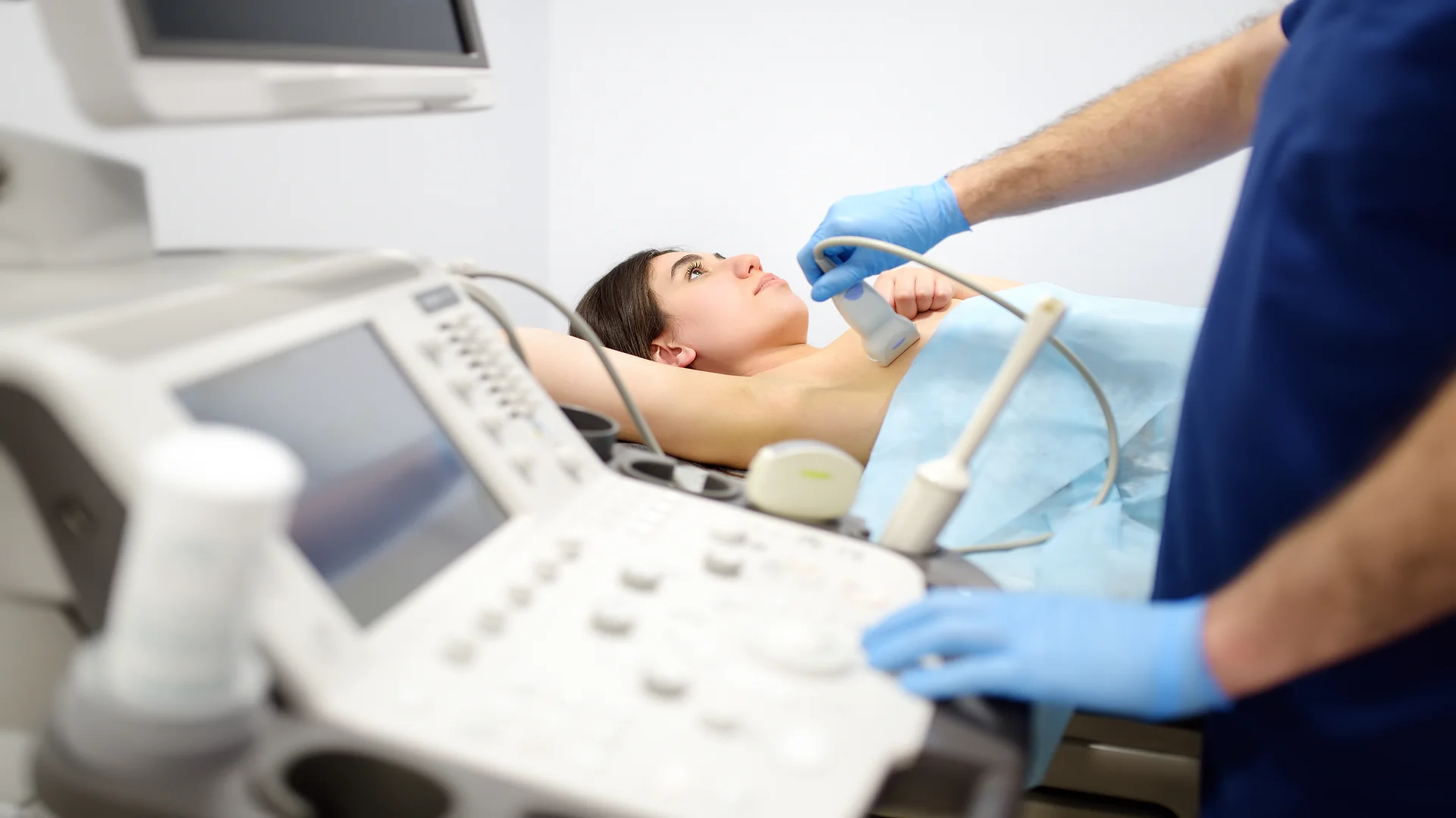
As already mentioned in the previous section of this breast ultrasound vs mammogram comparison, mammography is the standard screening method for most women. However, there are certain situations in which ultrasound offers an advantage, particularly when a breast abnormality needs closer examination.
According to RadiologyInfo’s guide to breast ultrasound, these situations include fluid-filled cysts, dense breast issues, breast biopsy, and breast lumps. Let’s learn more about each of them below.
1. Distinguishing Fluid-Filled Cysts From Solid Masses
One situation in which breast ultrasound is particularly useful is the evaluation of fluid-filled cysts. Mammography can show an abnormal mass, but it may not always clearly distinguish whether the lump contains fluid or consists of solid tissue. However, a breast ultrasound provides a closer look at its internal structure to show these findings apart.
A simple cyst typically appears as a fluid-filled structure, while a solid mass appears differently on the ultrasound image. This distinction helps determine whether further evaluation is needed.
2. Examining Areas Obscured by Dense Breast Tissue
Dense breast tissue can make abnormalities harder to identify on a mammogram because both dense tissue and many breast cancers appear white on X-ray images. As a result, when comparing mammography vs. ultrasound for dense breasts, ultrasound may be a more practical choice, as it can examine a different view of the breast and may reveal certain abnormalities hidden by overlapping dense tissue. This can make it a useful supplemental examination, particularly for women with dense breasts.
However, keep in mind that ultrasound does not detect every breast cancer and should not replace recommended mammography screening.
3. Guiding Breast Biopsy Procedures
When imaging reveals a suspicious breast abnormality, a radiologist may recommend a biopsy to determine whether the tissue is benign or cancerous. According to RadiologyInfo’s guide to ultrasound-guided breast biopsy, this technique is suitable when the abnormality is visible on ultrasound. During the procedure, real-time ultrasound images help the radiologist guide a needle into the targeted area and collect tissue samples for laboratory examination.
For more information about symptoms that may require medical attention, see our guide to the signs of cancer.
4. Evaluating Breast Lumps in Younger Women
Breast ultrasound is generally the initial imaging test for women younger than 30 who have a new palpable breast lump.
Younger women often have denser breast tissue, which can make certain abnormalities more difficult to assess on mammograms. For women aged 30–39, ultrasound and diagnostic mammography may both be appropriate, depending on the clinical findings.
What Mammography Can Detect More Reliably Than Breast Ultrasound
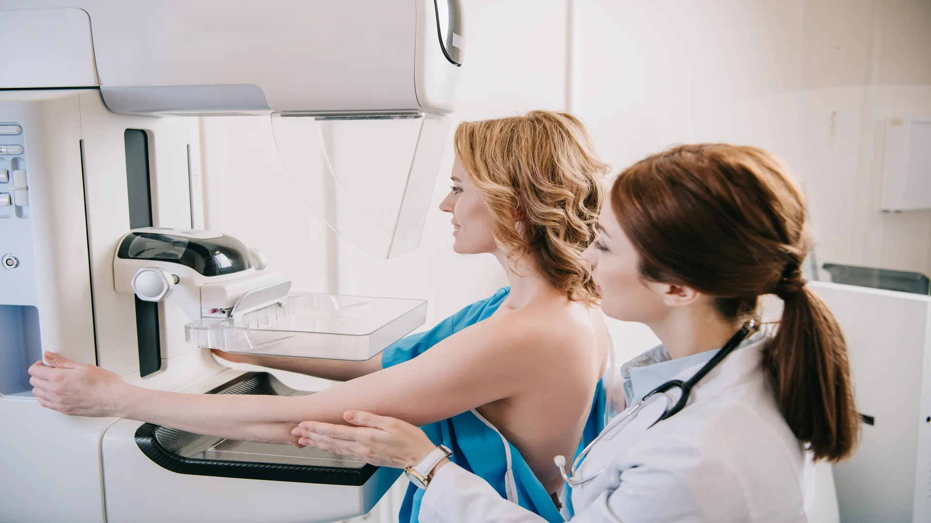
Unlike the situations discussed earlier, when breast ultrasound may offer an advantage, there are other findings for which mammography provides more useful information.
In a mammogram vs breast ultrasound comparison, clinicians may rely more on mammography to assess microcalcifications, architectural distortion, and asymmetries that may be less apparent on ultrasound alone. Medical studies identify these as important features of mammographic imaging.
1. Detecting Microcalcifications
Microcalcifications are tiny deposits of calcium within breast tissue, and mammography is more effective than ultrasound for detecting and characterizing them because it shows their pattern and distribution across the breast.
Ultrasound may still reveal some deposits, especially when they occur near or within a visible mass, and can provide complementary information. Certain microcalcification patterns can be associated with ductal carcinoma in situ (DCIS), a non-invasive breast cancer in which abnormal cells remain within the milk ducts.
2. Identifying Architectural Distortion and Asymmetry
Architectural distortion refers to a change in the normal arrangement of breast tissue. On a mammogram, tissue may appear drawn toward a point or crossed by radiating lines, even when no distinct mass is visible.
Another finding mammography can reveal is asymmetry, where an area of tissue looks different from the corresponding region in the opposite breast or from its expected appearance on other views. Radiologists compare the current mammogram with earlier images to see whether the finding is longstanding, new, or developing. If it persists or looks suspicious, additional mammographic views and targeted ultrasound may help clarify its significance.
Breast Ultrasound vs. Mammogram During Pregnancy and Breastfeeding
Pregnancy and breastfeeding are serious situations to consider when comparing breast ultrasound vs. mammography.
During these periods, hormonal changes can increase breast size, glandular tissue, and blood flow, and as a result, clinical examinations and image interpretation become more challenging. Also, some medical tests and treatments require additional precautions, and patients may wonder whether breast imaging should be avoided or whether one method is safer than the other.
The right choice depends on the patient’s age, breast cancer risk, and whether imaging is needed to investigate a symptom or for routine screening.
During pregnancy
Ultrasound is usually the first test for a new breast lump, focal pain, or suspicious nipple discharge. It uses no ionizing radiation and can examine the symptomatic area. However, if the findings are suspicious or ultrasound cannot explain the symptoms, diagnostic mammography may be added to detect calcifications and other abnormalities. Mammography is not prohibited, and indicated screening should continue according to age and risk.
During breastfeeding
The choice follows the usual age and symptom-based pathway rather than defaulting to ultrasound. For a new lump in women younger than 30, ultrasound is generally first. From age 30, diagnostic mammography or tomosynthesis is often combined with targeted ultrasound: mammography can show calcifications, while ultrasound examines the specific area. Routine screening also continues according to age and risk.
Before a mammogram during breastfeeding, nursing or pumping can reduce breast fullness, improve comfort, and potentially improve image quality. Furthermore, the X-rays used during mammography do not make breast milk radioactive, so there is no need to interrupt breastfeeding afterward. Most importantly, persistent lumps or other concerning breast changes should be evaluated promptly. Neither pregnancy nor breastfeeding should be a reason to delay medically necessary imaging.
Can Breast Ultrasound Replace a Mammogram?
No. Breast ultrasound cannot fully replace mammography because the two examinations deliver different types of information about breast tissue. Although they may sometimes be used to investigate the same abnormality, their imaging capabilities are not interchangeable. A finding that appears on ultrasound imaging may still require mammographic assessment, and vice versa.
Therefore, choosing one examination over the other is not simply a matter of preference; it requires considering what needs to be evaluated and which imaging method can provide the most reliable information. In some situations, using both methods offers a more complete assessment.
Breast Ultrasound vs Mammogram Age: Guidelines Compared
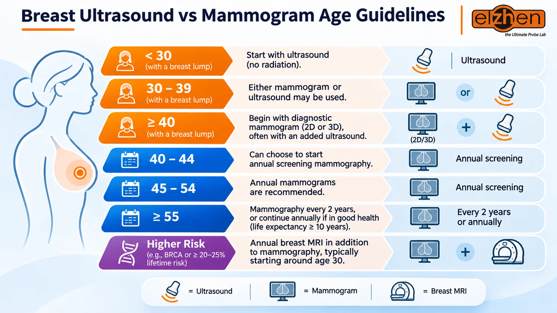
Now that you’ve explored the differences between breast ultrasound and mammography, you may still be wondering when each examination is recommended.
The recommended age and frequency of breast cancer screening depend on a person’s age and individual risk factors. Mammography is the primary screening method for most women at average risk, while ultrasound is generally used for diagnostic evaluation or supplemental screening in selected cases.
Here is a brief guide to when each examination may be appropriate.
- Women younger than 30 with a breast lump: Ultrasound is generally the initial diagnostic examination because it can evaluate the abnormality without ionizing radiation.
- Women aged 30–39 with a breast lump: Both diagnostic mammography and ultrasound may be appropriate initial examinations, depending on the clinical findings.
- Women aged 40 and older with a breast lump: Diagnostic mammography or 3D mammography is generally the initial examination, with targeted ultrasound often added for further evaluation.
- Women aged 40–44: According to the American Cancer Society (ACS), women can choose to begin annual screening mammography.
- Women aged 45–54: Annual mammograms are recommended for routine breast cancer screening.
- Women aged 55 and older: They can switch to mammography every two years or continue annual screening, provided they remain in good health and have an expected life expectancy of at least 10 years.
- Women at higher risk: Those with BRCA mutations or an estimated lifetime breast cancer risk of approximately 20–25% or higher may need annual breast MRI alongside mammography, typically starting around age 30.
A new breast lump, unusual nipple discharge, or other concerning changes should be evaluated promptly rather than waiting for the next routine screening appointment. Also, there is no universal starting age for routine breast ultrasound; its use depends on symptoms, imaging findings, breast density, and individual cancer risk.
Final Thoughts on Mammogram vs Breast Ultrasound
Throughout this mammogram vs. breast ultrasound comparison, we’ve explored how these examinations differ in their imaging technology, the findings they reveal, clinical applications, radiation exposure, etc.
Breast ultrasound is particularly useful for investigating specific breast abnormalities, evaluating lumps, and guiding biopsies, while mammography remains the primary screening method for most women at average risk and is more effective at detecting certain calcification patterns. Ultimately, neither examination is universally better; the right choice depends on the clinical situation and the patient’s individual needs.
Medical Sources and References
The medical information presented in this article is based on established medical guidelines, recognized healthcare organizations, and peer-reviewed research. The clinical explanations and recommendations have been researched and cross-checked against the following sources.
- American Cancer Society – Mammograms
Read the source - National Cancer Institute – Dense Breasts: Answers to Commonly Asked Questions
Read the source - American College of Radiology – ACR Appropriateness Criteria: Palpable Breast Masses
Read the source - Radiological Society of North America (RSNA) – Breast Imaging and Intervention During Pregnancy and Lactation
Read the source