CT scan vs ultrasound: Which imaging method is better for your medical condition?
When doctors need a closer look inside the body, choosing the right imaging test plays an important role not only in identifying a condition but also in guiding further medical decisions and treatment.
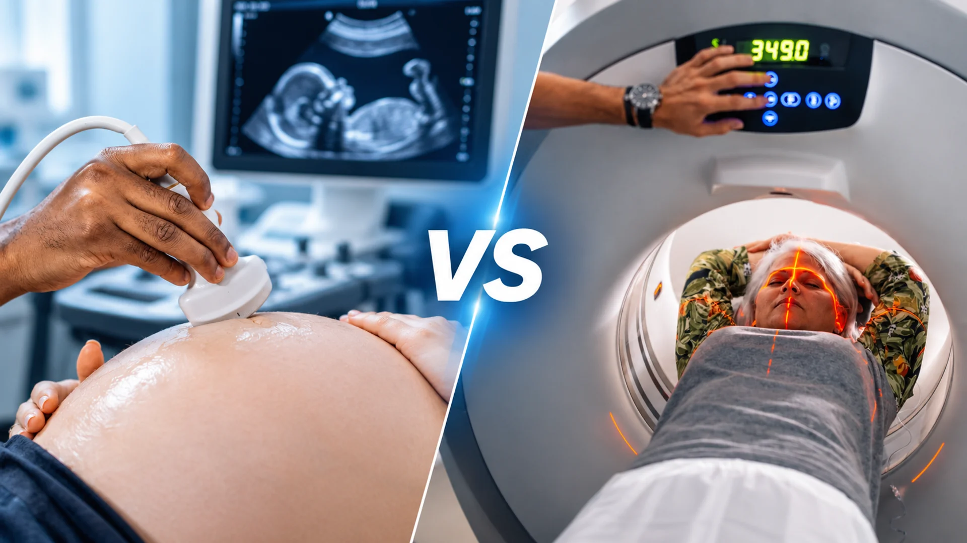
Ultrasound and CT scans are two widely used medical imaging methods, but they reveal different aspects of the body’s internal structures. So, when comparing Ultrasound vs CT scan, what makes one more suitable than the other?
Let’s explore how these imaging methods work, examine their strengths and limitations, and understand how doctors determine which examination is appropriate for different medical needs.
CT Scan vs Ultrasound Comparison at a Glance
The table below gives a quick comparison of CT scan vs ultrasound across several aspects, from how each scan works and its radiation exposure to image detail, typical uses, scan time, and cost.
| Feature | Ultrasound | CT Scans |
| Technology | Sends sound waves into the body and turns the returning echoes into an image | Rotates an X-ray beam around the body and uses a computer to build cross-sectional slices |
| Radiation | None | Yes, with the dose kept as low as is practical for each patient |
| What it Shows | Soft tissues, fluid-filled areas, and blood flow in a targeted area | Bones, lungs, organs, and blood vessels across a wider area |
| Limitation | Bowel gas, bone, body size, and scanning depth can reduce clarity | Lower radiation tolerance in some patients, and contrast dye may not suit everyone |
| Live viewing | Yes, so doctors can watch movement, heartbeat, and blood flow, and guide needles | Captures fast image sets but is not used for continuous viewing in routine scans |
| Common uses | Pregnancy checks, ovarian cysts, gallbladder problems, heart and vessel checks, and muscle or joint imaging | Injuries, suspected internal bleeding, lung disease, complex fractures, kidney stones in adults, and cancer assessment |
| Time and preparation | Usually around half an hour; some scans need fasting or a full bladder | Scanning takes only seconds to minutes, but the visit can run longer if fasting or contrast is needed |
| Cost and Access | Usually more affordable and available at the bedside with portable devices | Usually costlier and found mainly in hospitals and imaging centers |
This table provides a general overview of the differences between ultrasound and CT scans. To better understand how each of these types of medical imaging works and how they differ, read on: We begin with ultrasound, then move to CT scans, and finish with their seven key differences.
What Is an Ultrasound Scan?

To start our comparison of CT scan vs. ultrasound, let’s look at the technology behind ultrasound imaging.
An ultrasound scan, also known as sonography or ultrasonography, is a non-invasive medical imaging test that generates images without exposing patients to ionizing radiation. It is commonly used to evaluate internal organs, soft tissues, and blood flow.
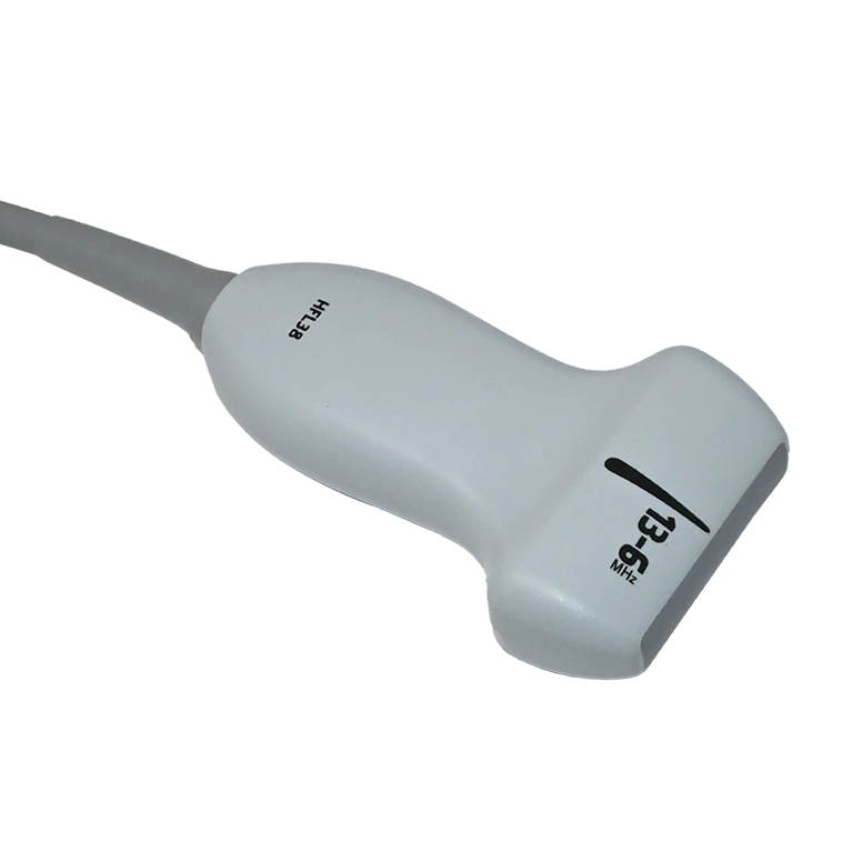
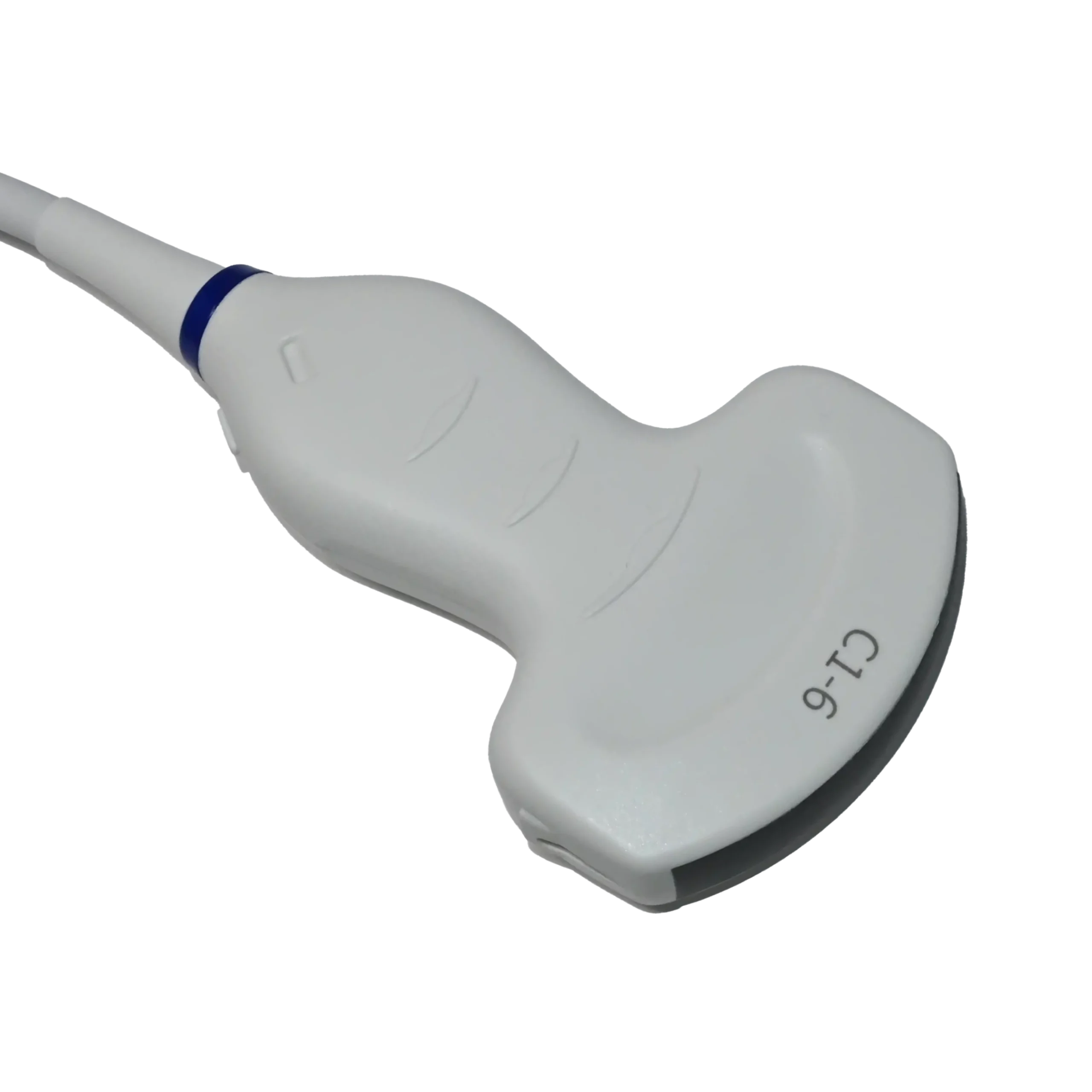
These examinations are performed using ultrasound machines equipped with different imaging technologies and features. Depending on their design and intended applications, these systems range from portable and handheld devices to advanced console-based machines. Ultrasound scans support various examinations, including abdominal, musculoskeletal (MSK), cardiac, and obstetric imaging, using specialized transducers. To explore their configurations and applications, read our guide to the different types of ultrasound machines.
Different Types of Ultrasound Machines
How Does an Ultrasound Scan Work?
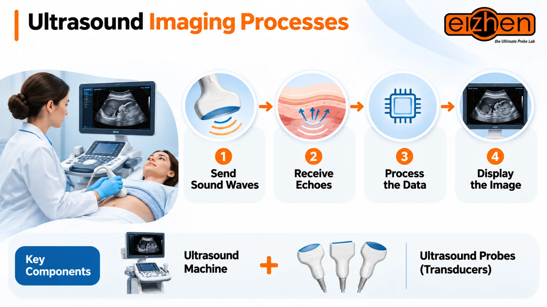
As mentioned earlier, ultrasound examinations can be performed using different types of ultrasound machines, each designed for specific clinical applications. However, the ultrasound machine is only part of the picture! It must be paired with a suitable ultrasound transducer to produce diagnostic images of the intended area.
The imaging process begins when the transducer sends high-frequency sound waves into the body and receives the echoes reflected by internal tissues; then the machine processes them into images on a monitor.
Similar to ultrasound machines, transducers come in different types based on their design, frequency, and the anatomical areas they examine.
Different Types of Ultrasound Probes
What Is a CT Scan (Computed Tomography)?
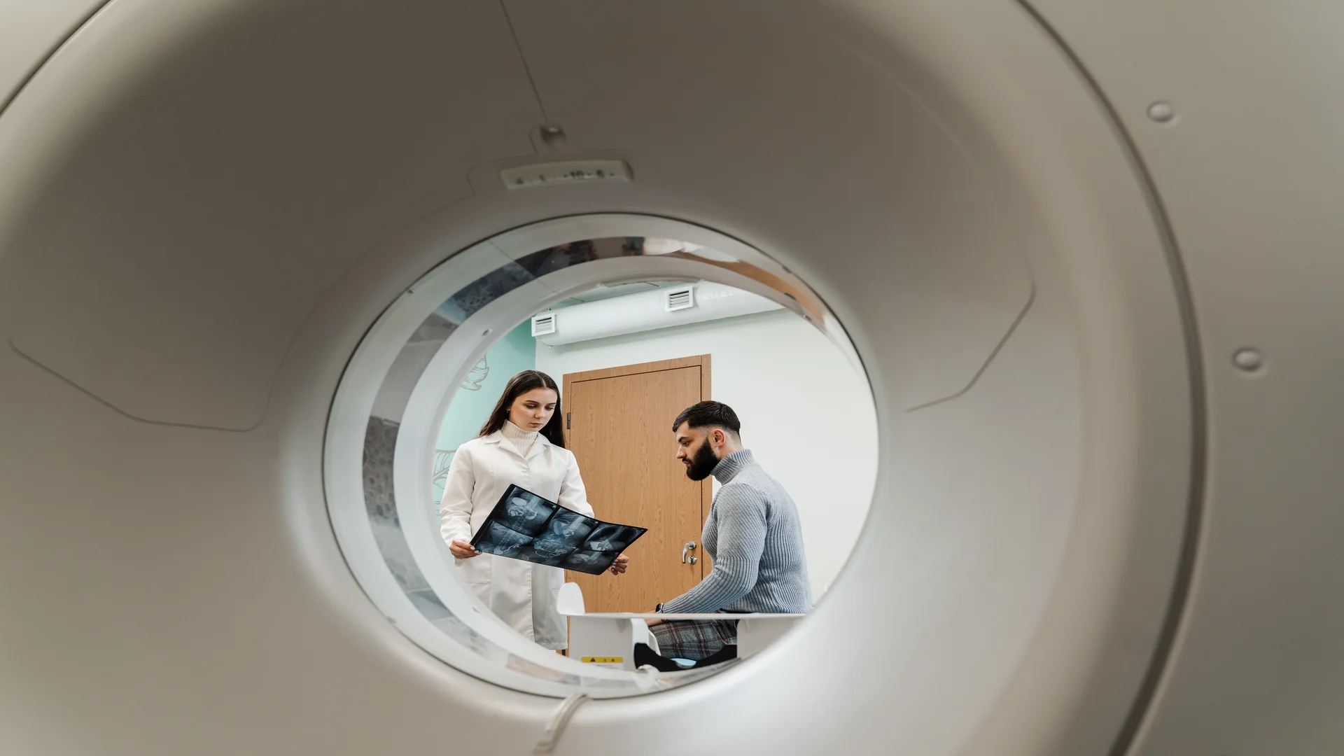
Now that we’ve explored ultrasound imaging, let’s look at the other side of the CT scan vs ultrasound comparison: Computed Tomography. Understanding how CT scans work will help us better understand the differences between these two imaging methods.
A CT scan, short for computed tomography, is a medical imaging examination that uses X-rays and computer technology to create detailed images of the inside of the body. Unlike conventional X-rays, which produce two-dimensional images, CT scans capture multiple images from different angles and combine them to create cross-sectional views of internal structures. These images allow doctors to examine organs, bones, blood vessels, and other tissues in greater detail.
Although the examination itself is non-invasive, some CT procedures require an injection of contrast material, usually through an intravenous (IV) line, to make certain structures and abnormalities easier to see.
How Does CT Scan Work?
A CT scanner uses X-rays, a form of electromagnetic radiation that can pass through the body and help create images of internal structures.
As shown in the image below, during the examination, the patient lies on a movable table that passes through the scanner while an X-ray tube rotates around the body, capturing information from different angles.
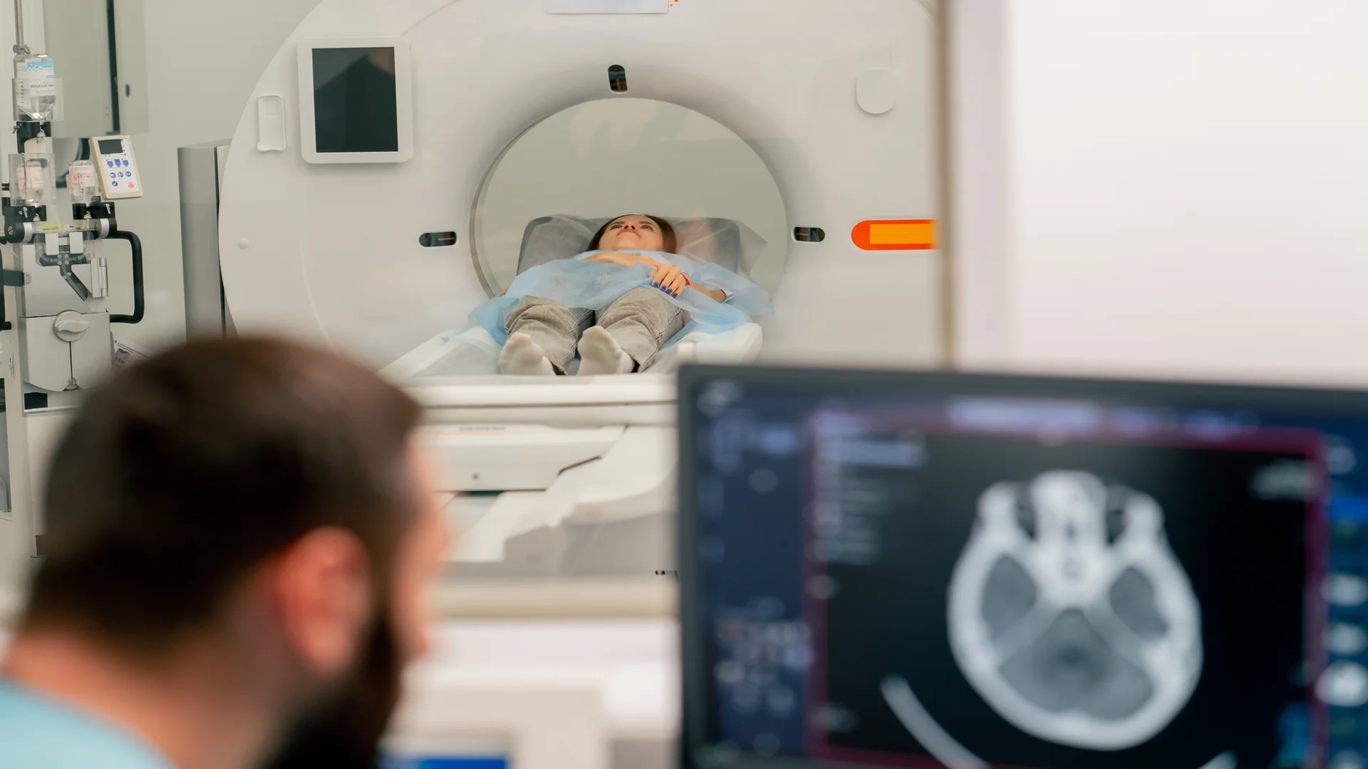
As X-rays travel through the body, different tissues weaken the X-ray beam to varying degrees, allowing the scanner to distinguish between structures such as bones and soft tissues. A computer processes the collected information to reconstruct detailed cross-sectional images, also known as slices, of the examined area.
What Are the Types of CT Scans?
Similar to ultrasound scans, CT examinations can target different anatomical areas depending on the patient’s symptoms and the diagnostic information needed. Some of the most common examinations include:
- Head CT: Examines the brain and skull, often to investigate head injuries, bleeding, or suspected stroke.
- Chest CT: Evaluates the lungs, heart, and other structures within the chest.
- Abdominal and Pelvic CT: Examines organs and tissues in the abdomen and pelvis to investigate conditions such as infections, inflammation, and tumors.
- Spinal CT: Provides detailed images of the spine, particularly when fractures or other abnormalities are suspected.
- CT Angiography (CTA): Uses contrast material to examine blood vessels and identify conditions such as narrowing, blockages, or aneurysms.
- Cardiac CT: Evaluates the heart and coronary arteries, including through coronary calcium scoring and coronary CT angiography.
CT Scan vs Ultrasound: 7 Key Differences
Although ultrasound and CT are both widely used medical imaging techniques, they serve different purposes. A scan that captures blood flow in real time (Ultrasound) may answer a very different diagnostic question from one that provides detailed cross-sectional views of the body (CT Scan).
So, when comparing CT scan vs ultrasound, there’s more to consider than just how each technology works. Factors such as radiation exposure, image detail, clinical applications, scan time, and patient experience can all influence which method is more suitable for a particular medical condition.
Let’s take a closer look at seven key differences between these two imaging methods.
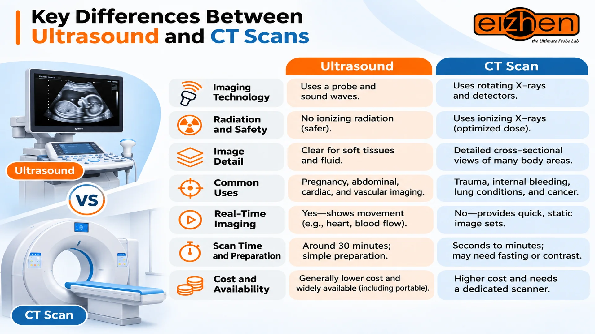
1. Imaging Technology: How Does Each Scan Produce Images?
The main difference between Ultrasound and CT scan lies in how they receive and process information to create images:
- Ultrasound: Uses a handheld transducer (probe), such as a phased array probe or linear transducer, to transmit and receive sound waves and then produce images.
- CT scan: Uses an X-ray tube and detectors that rotate around the patient to collect information from different angles, which a computer reconstructs into cross-sectional images.
2. Radiation and Safety: How Do the Risks Differ?
Radiation exposure is an important safety consideration when comparing CT scan vs ultrasound. The two techniques differ in the type of energy they use and the potential risks associated with an examination:
- Ultrasound: Uses non-ionizing sound waves and does not expose patients to ionizing radiation. As a result, it could be the preferred imaging method for fetal monitoring and many follow-up examinations.
- CT scan: Uses ionizing X-rays, so clinicians optimize the radiation dose according to the patient’s needs and the diagnostic purpose. Some examinations also require iodine-based contrast material, which may involve additional considerations for patients with certain allergies or impaired kidney function.
3. Image Detail: What Can Each Scan Reveal?
Neither technique provides better images for every anatomical structure. Their imaging capabilities depend on the tissues being examined and the diagnostic information required:
- Ultrasound: Produces detailed images of many superficial soft tissues, fluid-filled structures, and blood flow. However, visualization can be limited by bowel gas, bone, examination depth, patient body habitus, and operator technique.
- CT scan: Provides detailed cross-sectional views of broader anatomical areas, including bones, lungs, organs, and blood vessels.
4. What Is Each Imaging Method Best For?
The choice between ultrasound and CT often depends on the suspected condition, the urgency of the diagnosis, and the anatomical area being evaluated. Their common clinical applications include:
- Ultrasound: Pregnancy monitoring, abdominal ultrasound imaging, cardiac and vascular assessment, and musculoskeletal ultrasound.
- CT scan: Traumatic injuries, suspected internal bleeding, lung conditions, complex fractures, and certain cancer evaluations.
For some conditions, one method may be preferred as the initial examination, while the other may be recommended when additional diagnostic information is needed.
As we continue our comparison of CT scan vs ultrasound, we’ll take a closer look at medical conditions where both imaging methods may be used.
5. Real-Time Imaging: Can Doctors Observe Movement?
A major advantage of ultrasound imaging is its ability to display images continuously during an examination. This is particularly useful when clinicians need to assess movement or observe changes as they happen:
- Ultrasound: Allows clinicians to observe organ movement and heart activity, assess blood flow using Doppler imaging, and guide procedures such as biopsies and fluid drainage in real time.
- CT scan: Captures rapid sets of images rather than providing continuous visualization during routine examinations.
6. Scan Time and Preparation: What Should You Expect?
The time required for each examination and the preparation involved depend on the body area being scanned and the imaging protocol:
- Ultrasound: Examinations often take around 30 minutes, although some are shorter or longer. Preparation may involve fasting before an abdominal scan or drinking water to ensure a full bladder for certain pelvic examinations.
- CT scan: Image acquisition may take only seconds to minutes, although the overall appointment can be longer when preparation or contrast administration is required. Some examinations involve fasting or drinking contrast material beforehand.
7. Cost and Availability: Which Is More Accessible?
Cost is another practical factor when comparing CT scan vs ultrasound. Differences in equipment, infrastructure, and clinical settings influence how easily each examination can be accessed:
- Ultrasound: Examinations are generally less expensive, and portable and handheld systems make bedside imaging possible in hospitals and clinics.
- CT scan: Usually involves higher equipment and operating costs and requires dedicated scanners, which are typically available in hospitals and specialized imaging centers.
All in all, actual examination prices and availability vary depending on the facility, location, and type of scan.
When Is Ultrasound Better Than CT?
In the previous sections, we compared CT scan vs ultrasound in terms of how they work, image quality, safety, and other key differences.
Now, let’s look at when one imaging method may be more suitable than the other. Two good examples are ovarian cysts and gallbladder conditions, where ultrasound is generally the preferred initial examination.
Ovarian Cysts: CT Scan vs Ultrasound
When evaluating an ovarian cyst, ultrasound is usually preferred over CT for the initial examination. Both transvaginal and transabdominal ultrasound can show the cyst’s size, structure, and whether it contains fluid or solid components. Doppler imaging can also assess blood flow, helping doctors evaluate the surrounding ovarian tissue.
Gallbladder CT Scan vs Ultrasound
For suspected gallstones or gallbladder inflammation, ultrasound is generally the first-choice imaging method. It can identify gallstones, gallbladder wall thickening, and fluid around the gallbladder. As a result, it would be a better choice for evaluating suspected cholecystitis.
CT may be used when ultrasound findings are inconclusive or when doctors need to investigate complications or other possible causes of abdominal pain.
When Is a CT Scan Better Than Ultrasound?
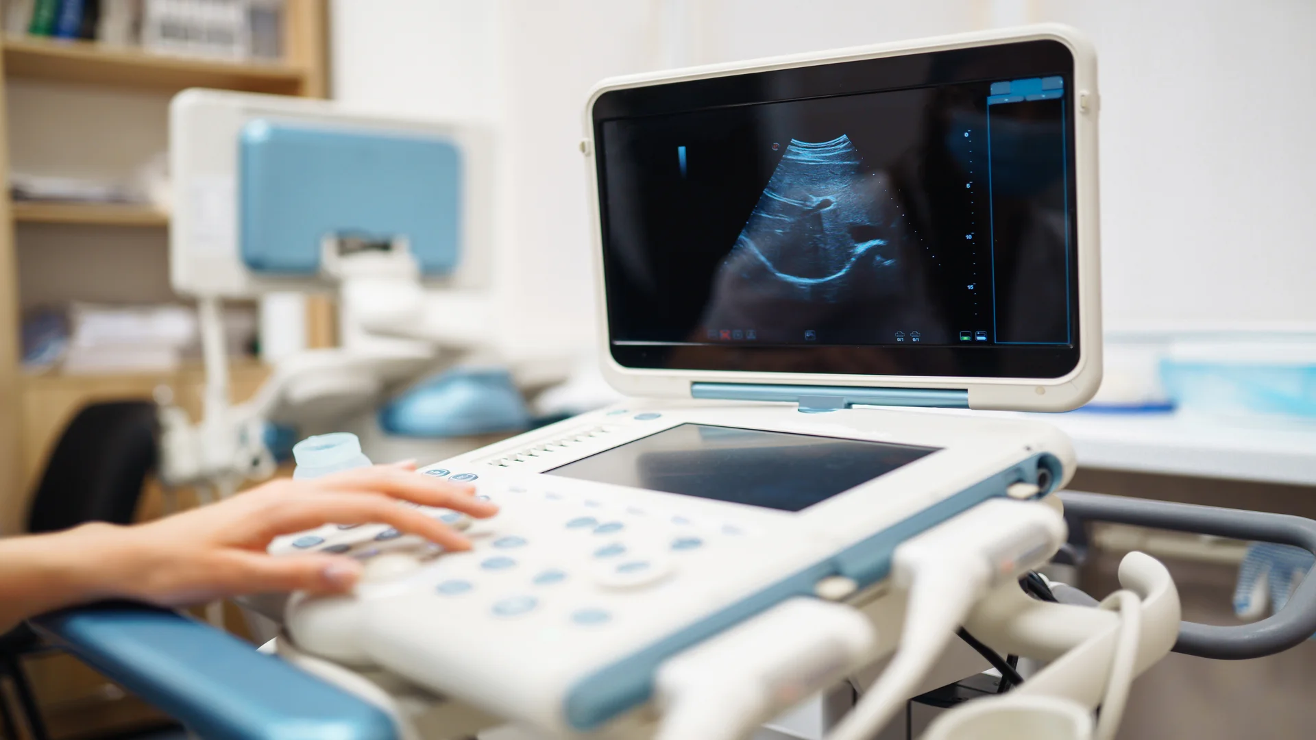
However, ultrasound is not always the preferred option. For example, when it comes to CT scan vs ultrasound for kidney stones, CT scans may provide more reliable diagnostic information than ultrasound.
Medically speaking, a non-contrast CT scan is generally more sensitive for detecting urinary stones, including small stones in the ureter, and can show their exact size and location.
Ultrasound can identify kidney swelling caused by an obstruction, but it may miss smaller stones. Nevertheless, ultrasound is often preferred initially for children and pregnant patients to avoid radiation exposure.
Can You Need Both an Ultrasound and a CT Scan?
Can your doctor recommend a CT scan after an ultrasound, or an ultrasound after a CT scan?
Yes. As explained earlier in this CT scan vs ultrasound comparison, these methods provide different but complementary information. For example, in an abdominal ultrasound vs CT scan evaluation, ultrasound may be performed first to examine a specific organ, such as for suspected appendicitis. If the findings are inconclusive or suggest a more complex condition, a CT scan can provide a broader view of the situation.
The reverse is also possible. If a CT scan reveals an unclear abnormality, a targeted ultrasound may help doctors examine it more closely. For instance, ultrasound can help distinguish fluid-filled cysts from solid lesions in certain organs. Ultimately, the decision to use one or both methods depends on the suspected condition and the additional information needed for diagnosis.
Conclusion
In this CT scan vs ultrasound comparison, we’ve explored the key differences, applications, and limitations of both imaging methods. Ultrasound is a non-invasive technique that uses sound waves without ionizing radiation, although its imaging capabilities can be limited by bowel gas, bone, and examination depth.
CT scans provide detailed cross-sectional images, making them particularly useful for evaluating complex conditions, injuries, and internal structures. Ultimately, neither method is universally better; they serve different diagnostic purposes and can sometimes complement each other to help doctors reach a more accurate diagnosis.