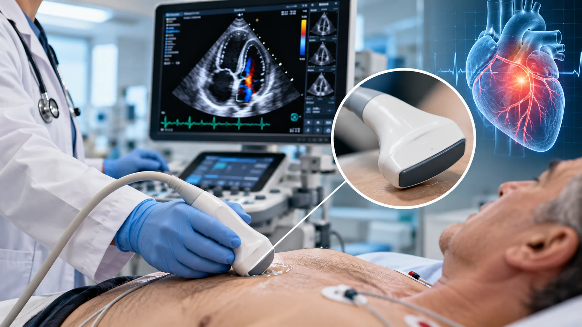
With cardiovascular disease accounting for nearly one in three deaths worldwide, examining the heart and blood vessels is an important part of cardiovascular care.
In cardiology, ultrasound is widely used to support this assessment, offering real-time imaging without ionizing radiation. Depending on what needs to be examined, clinicians may use different transducers for echocardiography or vascular ultrasound.
This article explores these applications and probe types to help clarify which cardiovascular transducers are suited to different imaging needs.
From Cardiology to Ultrasound Assessment
Cardiology is a branch of medicine focused on the heart and cardiovascular system, including diagnosing, managing, and preventing conditions affecting the heart and blood vessels. It covers a broad range of cardiovascular conditions and clinical areas, including coronary artery disease, heart rhythm disorders, heart failure, valvular heart disease, and vascular disease.
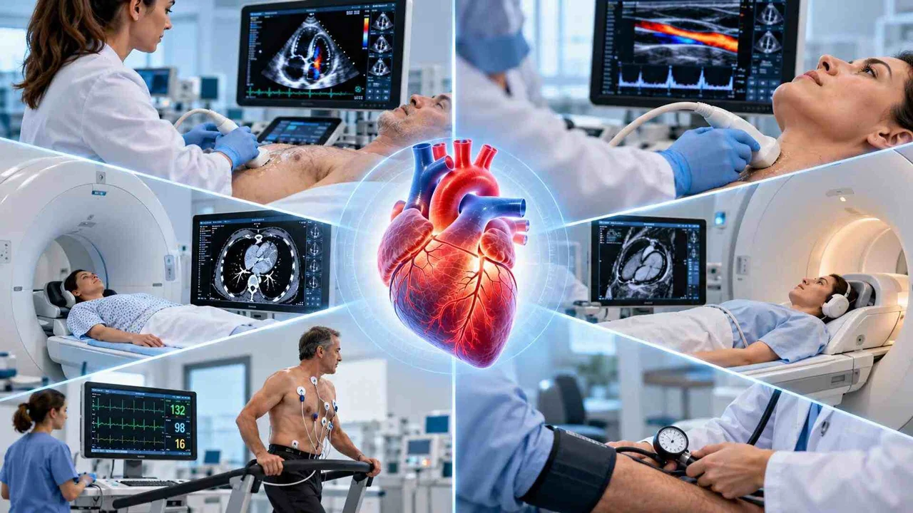
Because these conditions can affect different parts and functions of the cardiovascular system, cardiology relies on a variety of diagnostic and assessment methods. Depending on the patient’s condition and the information needed, these may include clinical examination, electrocardiography (ECG/EKG), cardiac stress testing, echocardiography, cardiac CT, cardiac MRI, vascular ultrasound, and other cardiovascular tests.
Among these methods, cardiovascular ultrasound plays an important role in assessing both the heart and blood vessels using different types of new or refurbished ultrasound probes. The following section takes a closer look at this type of examination and its role within cardiology.
How Cardiovascular Ultrasound Is Performed in Cardiology
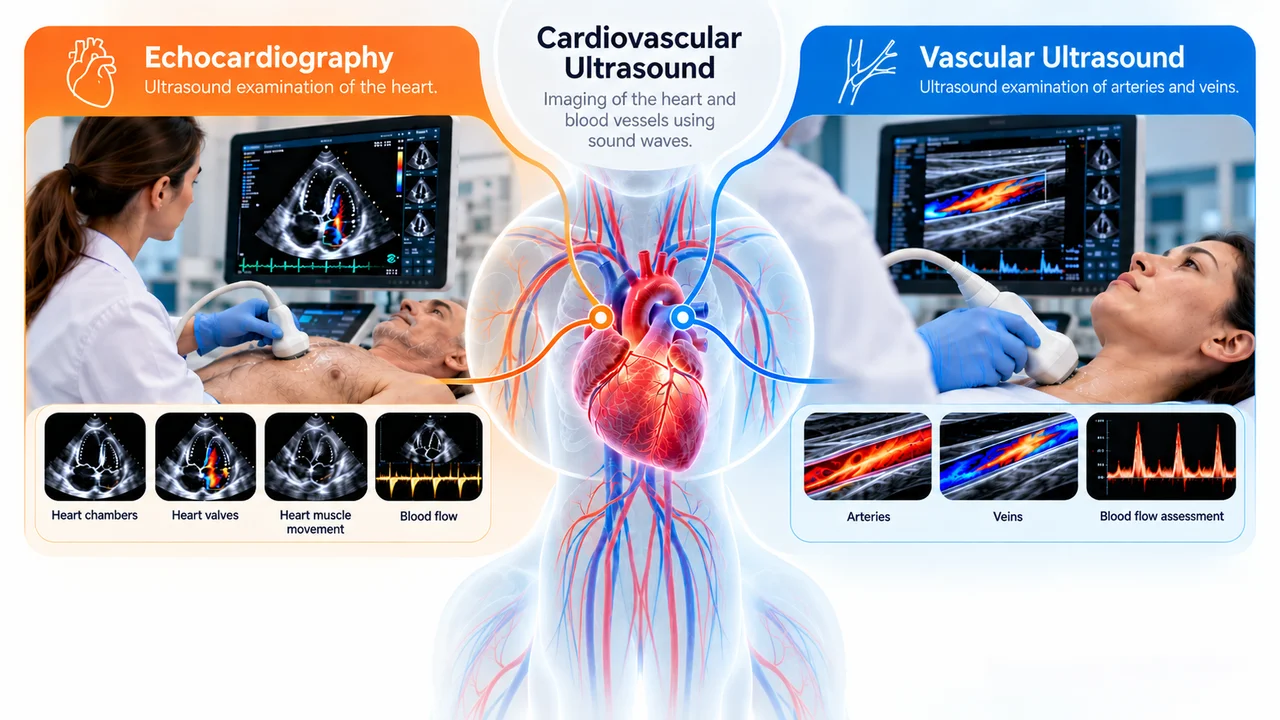
As mentioned above, cardiovascular ultrasound is one of the imaging methods used in cardiology, using sound waves to examine different parts of the cardiovascular system, including the heart and blood vessels. Since these examinations may target different parts of this system, cardiovascular ultrasound can take different forms depending on what needs to be assessed, including echocardiography and vascular ultrasound.
In both cases, ultrasound systems and transducers provide real-time imaging without ionizing radiation, making it a useful and generally safe method for evaluating cardiovascular structures, function, and blood flow. Below, we look more closely at how each of these examinations contributes to cardiovascular assessment.
Echocardiography
When cardiovascular ultrasound is focused on the heart, it is performed through echocardiography. This examination allows clinicians to assess cardiac structures and function, including the heart chambers, valves, muscle movement, and blood flow.
For example, a patient with suspected heart valve disease may undergo a transthoracic echocardiogram (TTE) to evaluate the condition and function of the valves. During TTE, the transducer is positioned at different areas of the chest to obtain multiple views of the heart, allowing clinicians to examine the valves alongside other cardiac structures and assess how the heart is functioning.
Vascular Ultrasound
While echocardiography focuses on the heart, vascular ultrasound extends cardiovascular assessment to the arteries and veins. It provides real-time images of these vessels and allows clinicians to evaluate blood flow, which can help identify abnormalities such as arterial narrowing, blockage, or venous blood clots.
For example, a patient with symptoms such as sudden weakness or numbness on one side of the body or temporary difficulty speaking may need further evaluation for possible vascular disease. If carotid artery narrowing is suspected, vascular ultrasound can be used to visualize the carotid arteries and assess blood flow through them. Doppler techniques are often incorporated into the examination to provide additional information about the direction and characteristics of blood flow.
Types of Cardiovascular Ultrasound Probes Used in Cardiology
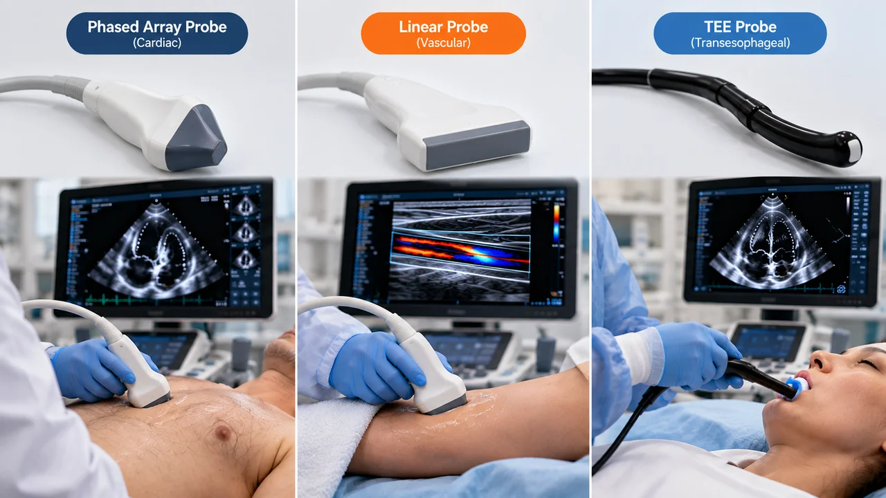
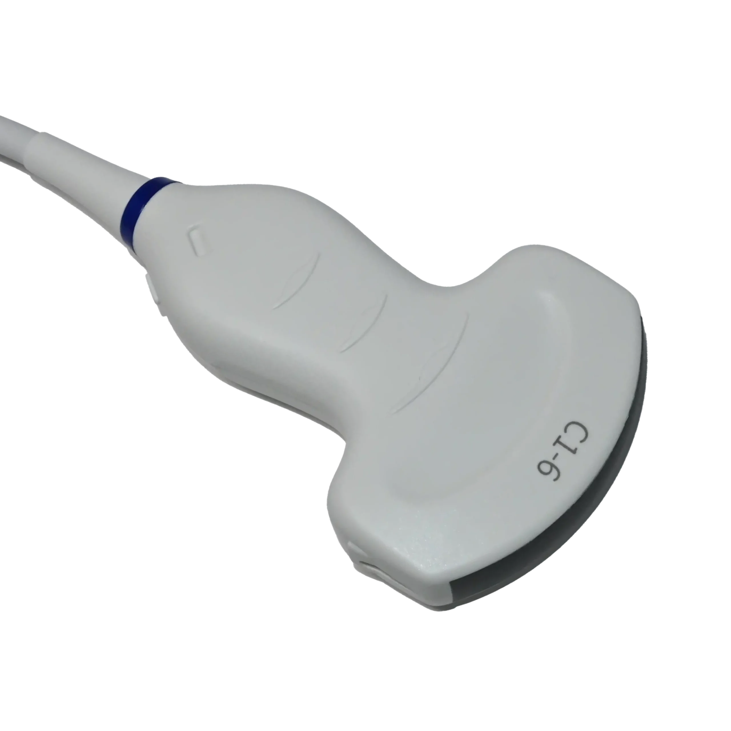
As discussed above, ultrasound supports a range of cardiology applications, particularly cardiac and vascular imaging. However, not every cardiovascular examination uses the same probe; clinicians select different transducer types based on the anatomy being examined, the imaging approach, required depth, and patient needs. Below are some of the main transducers used across cardiovascular ultrasound.
Phased Array Transducers
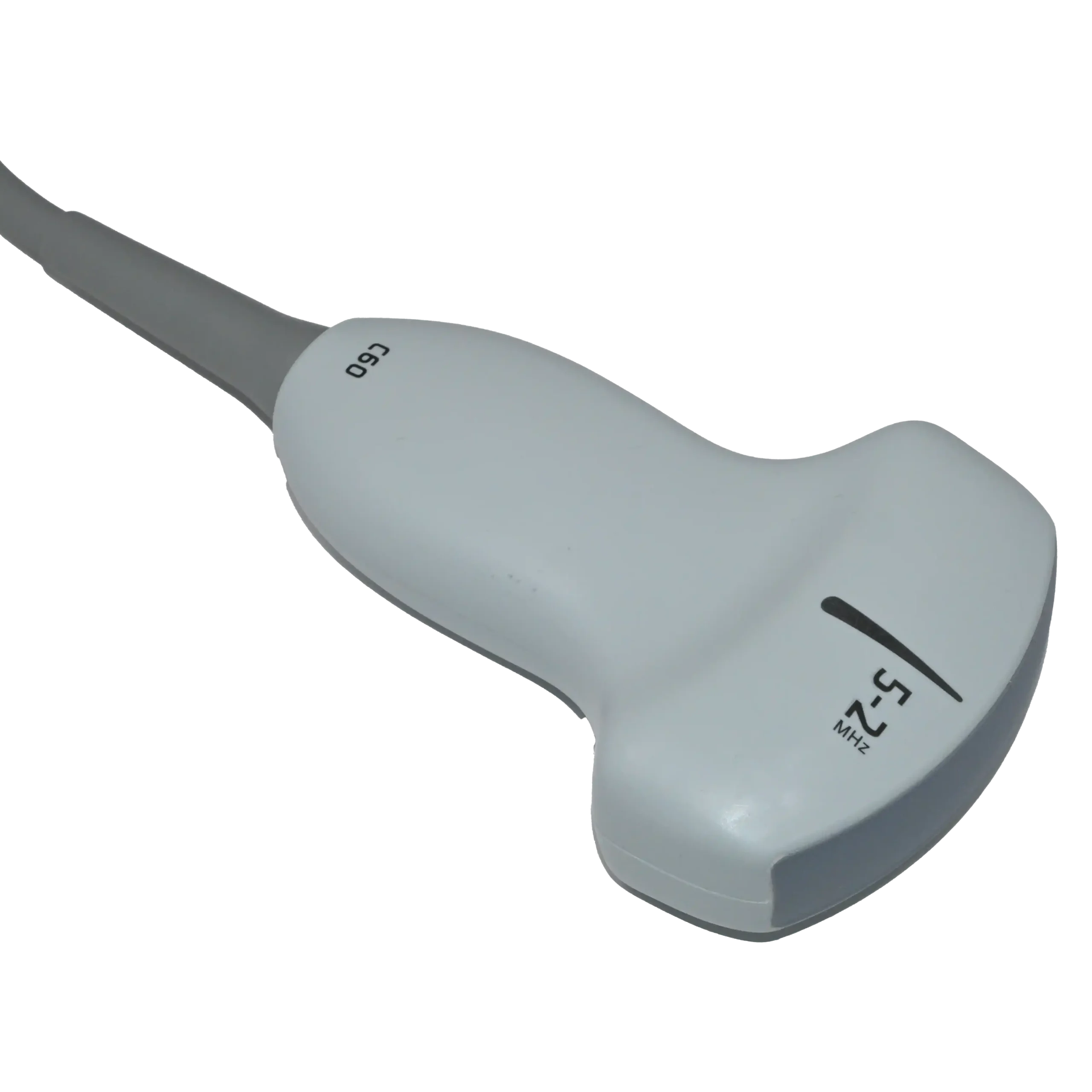
Phased array transducers are widely used in echocardiography, particularly for imaging the heart through the chest. Their small footprint and sector-shaped field of view make them well suited for imaging through the spaces between the ribs. We have explained how this design works and where it is used in more detail in our article about phased array transducers.
Linear Array Transducers
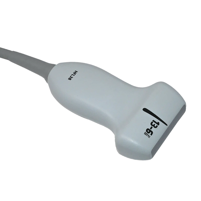
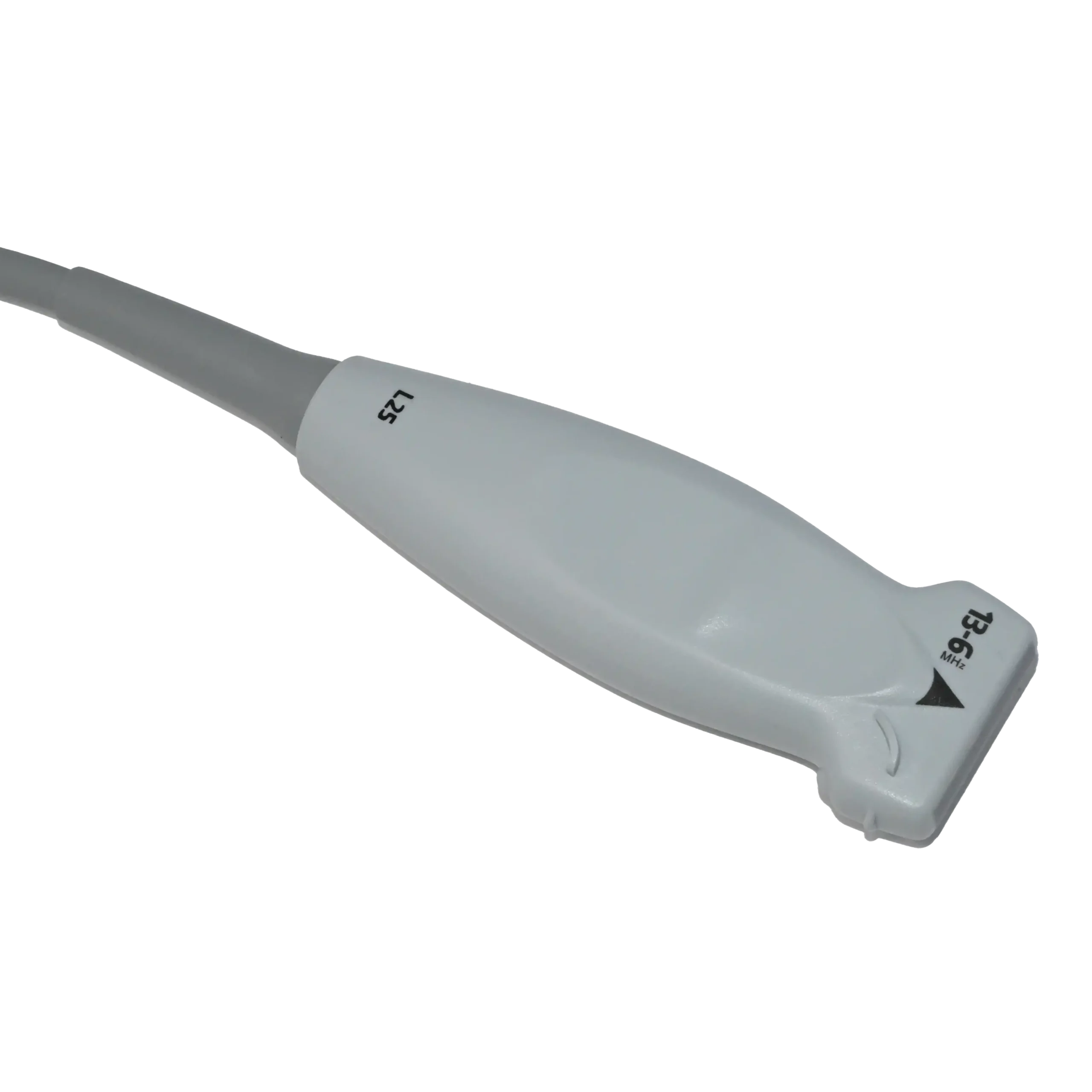
While phased array probes are mainly associated with cardiac imaging, linear array transducers are commonly used for superficial vascular examinations. Their straight array produces a rectangular image with good resolution at shallow depths, making them suitable for examining superficial arteries and veins. You can learn more about this design and its other medical applications in our article on linear transducers.
TTE and TEE in Echocardiography
In the previous sections, we discussed specific transducer array designs used in echocardiography and cardiovascular ultrasound. Transthoracic echocardiography (TTE) and transesophageal echocardiography (TEE), however, are not array types but two approaches to cardiac imaging that determine how and where the probe is used.
TTE is performed through the chest and typically uses a phased array probe, while TEE is performed from within the esophagus using a specialized transesophageal probe, which may use phased-array or matrix-array technology. To learn more about TEE imaging and how it works, read our article “What Is a TEE Transducer?”.
Phased Array vs. Linear vs. TEE Probes at a Glance
The table below helps you better understand the key differences between common ultrasound transducers used in cardiology and cardiovascular examinations.
| Feature | Phased Array Probe | Linear Probe | TEE Probe |
| Classification | An ultrasound transducer array type | An ultrasound transducer array type | A specialized probe design used for a specific echocardiographic approach |
| Typical image shape | Sector-shaped image that expands with depth | Rectangular image | Typically sector-based cardiac views; exact imaging capabilities depend on the probe technology |
| Probe placement | Placed on the chest at different acoustic windows | Placed on the skin over the vessel or structure being examined | Through the mouth and positioned in the esophagus |
| Typical imaging depth | Suitable for imaging deeper structures such as the heart | Best suited to relatively superficial structures; lower-frequency linear probes can reach | Provides close access to the heart from the esophagus, reducing the distance between the transducer and many cardiac structures |
| Main role in cardiology | Cardiac imaging and transthoracic echocardiography (TTE) | Vascular imaging, including assessment of superficial arteries and veins | Transesophageal echocardiography (TEE) for detailed cardiac and structural imaging |
| Common cardiovascular targets | Heart chambers, valves, ventricular function, and blood flow | Superficial arteries, veins, vessel walls, and surrounding tissues | Heart valves, atria, left atrial appendage, septal structures, and other cardiac anatomy |
| Key advantage | Combines a small contact area with the depth and sector view needed for cardiac imaging | Provides high spatial resolution for superficial vascular structures | Can provide detailed views of cardiac structures that may be difficult to visualize adequately with transthoracic imaging |
| Main limitation | Generally provides less near-field detail than a high-frequency linear probe | Its rectangular footprint and typical frequency range make it less suited to routine transthoracic cardiac imaging | More invasive than TTE and requires appropriate patient preparation, clinical expertise, and monitoring |
Which Cardiovascular Probe Is Best in Cardiology?
With several cardiovascular probe types used across cardiac and vascular imaging, a common question is which is best for cardiology. However, no single cardiovascular ultrasound probe is best for every examination. The right choice depends mainly on what needs to be examined, the imaging approach, the required depth, and the patient.
For example, when a patient needs an ultrasound examination of superficial arteries or veins, a linear array transducer is often suitable because it provides high-resolution imaging at shallow depths.
For transthoracic echocardiography, however, a phased array probe is typically preferred because its small footprint helps clinicians obtain cardiac views through the spaces between the ribs. When closer views of the heart are required through a transesophageal approach, a specialized TEE probe is used instead.
Therefore, choosing the best cardiovascular probe starts with the intended examination, not a one-size-fits-all transducer. If you are looking specifically for heart imaging equipment, you can explore our available cardiac ultrasound probes, with options for different ultrasound systems and cardiac imaging requirements.
What to Expect During a Cardiovascular Ultrasound Examination
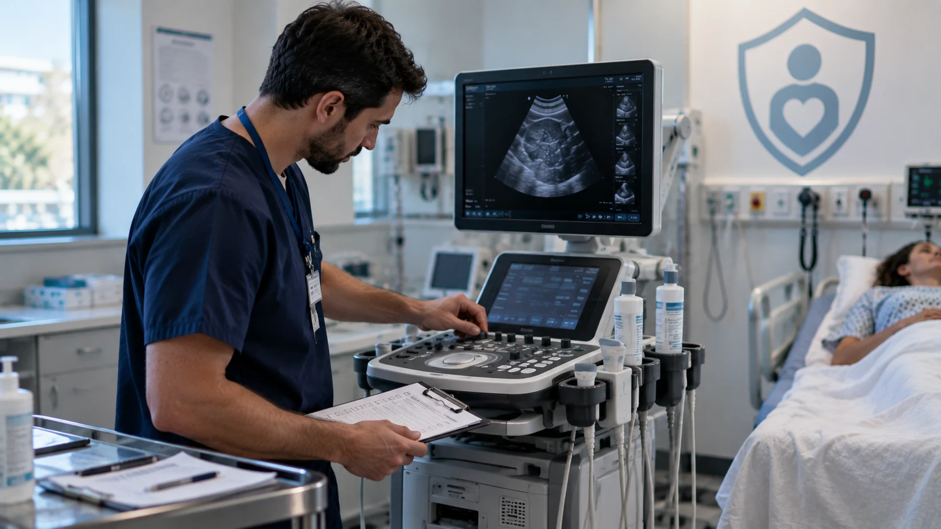
A qualified healthcare professional performs a cardiovascular ultrasound examination using a transducer suited to the specific imaging task and an ultrasound system that supports it. The setting can vary by examination and may include a hospital, clinic, imaging center, or medical office. During the scan, the transducer is placed or used according to the cardiovascular area being examined and the type of images required.
What happens before and after the examination also depends on the ultrasound approach. Standard transthoracic echocardiography (TTE) and many vascular ultrasound examinations generally involve minimal preparation, with patients usually able to continue their usual routine afterward.
Transesophageal echocardiography (TEE), however, involves a different process because the probe is passed into the esophagus to obtain images of the heart. Patients are typically instructed to avoid eating or drinking for a specified period beforehand, and sedation is commonly used during the procedure. Some observation may also be needed afterward before the patient leaves. Because requirements can differ between examinations and healthcare facilities, patients should follow the instructions given by their medical team, including any guidance related to medications.
Choosing the Right Cardiovascular Probe for Your Cardiology Department
Throughout this article, we have taken a closer look at the role of ultrasound transducers in cardiology, the different probes used for echocardiography and vascular imaging, and a common question: which cardiovascular ultrasound probe is best? As we have seen, there is no single answer. Each probe has its own imaging capabilities, and the right choice depends on the examination, clinical requirements, patient needs, and compatibility with the ultrasound system.
If you are looking for a cardiovascular probe for a hospital, clinic, or cardiology department and are unsure which option fits your needs, Elzhen can help. Our team can help you find a compatible cardiac ultrasound probe based on your ultrasound system and imaging requirements. Contact Elzhen to discuss the probe you are looking for and check available options.