How can doctors see inside the human body without performing surgery?
Various types of medical imaging make this possible through different technologies that reveal internal organs, tissues, bones, and blood vessels. Each technique provides different information to support diagnosis and treatment.
Keep scrolling to understand how these imaging techniques work and what makes each type of medical scan unique, as well as how they are used in modern healthcare.
What Is Medical Imaging?
You may have experienced this during a doctor’s visit: after a physical examination, your doctor recommends an additional test, such as an ultrasound, X-ray, CT scan, or MRI, to investigate your condition further.
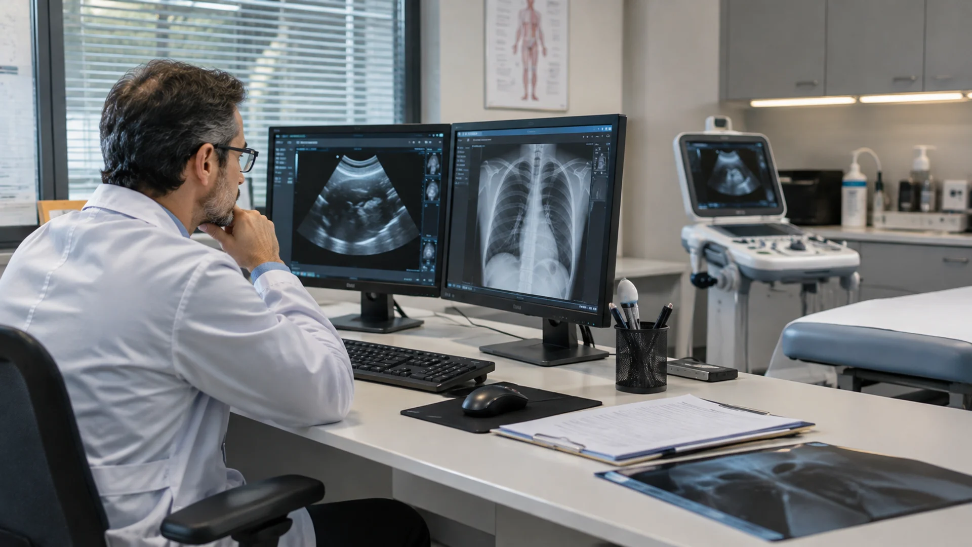
These types of medical imaging allow healthcare professionals to examine different organs and tissues of the body, each providing a different type of information.
The choice of examination depends on factors such as the suspected condition, the body area being assessed, and the clinical information needed. Clinicians then interpret imaging findings alongside the patient’s symptoms, medical history, physical examination, and other test results to establish an accurate diagnosis and guide further care.
6 Types of Medical Imaging or Medical Scans
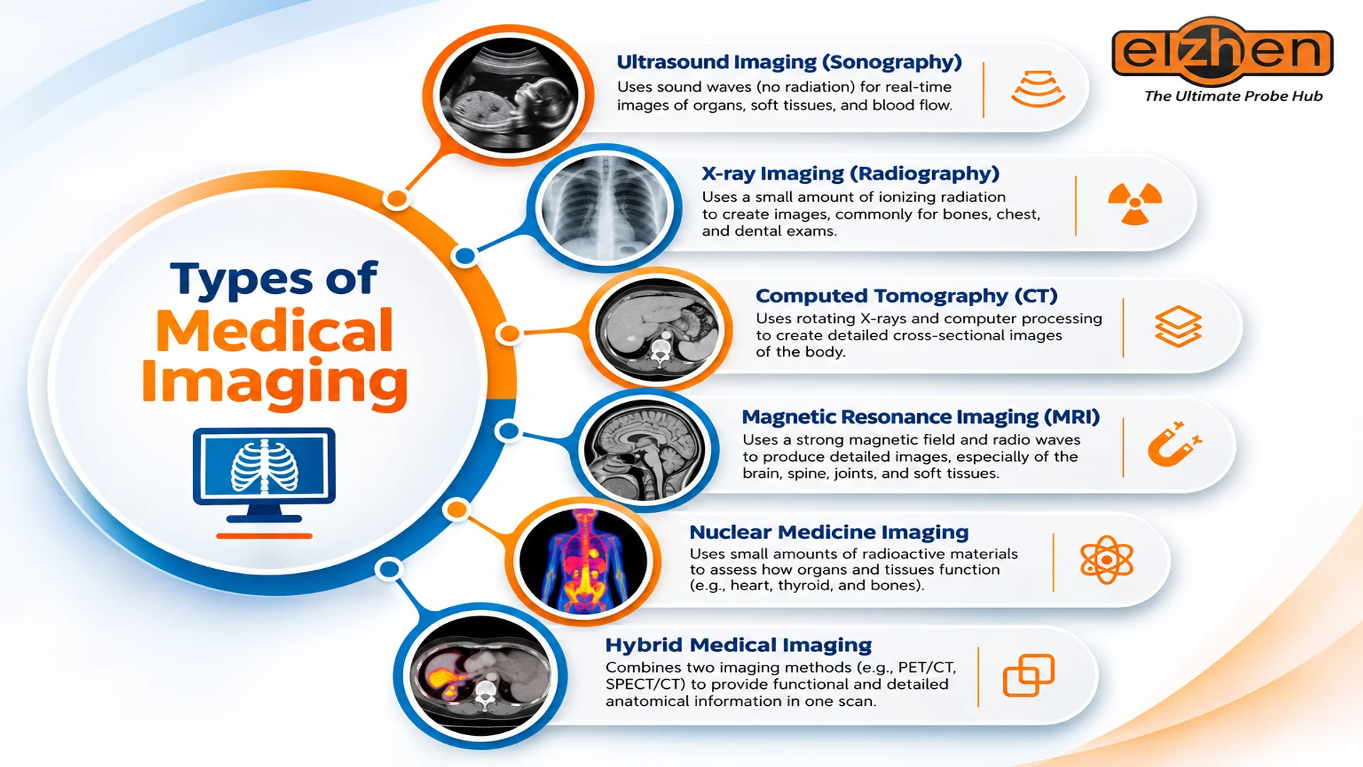
The most common types of medical imaging, also known as diagnostic imaging tests or types of medical scans, include ultrasound, X-ray, computed tomography (CT), magnetic resonance imaging (MRI), and nuclear medicine imaging. Each method uses different technology to examine the body and provide information for clinical assessment.
In the following sections, we’ll take a closer look at how each method works and its common medical applications, starting with ultrasound systems.
Ultrasound Imaging (Sonography)
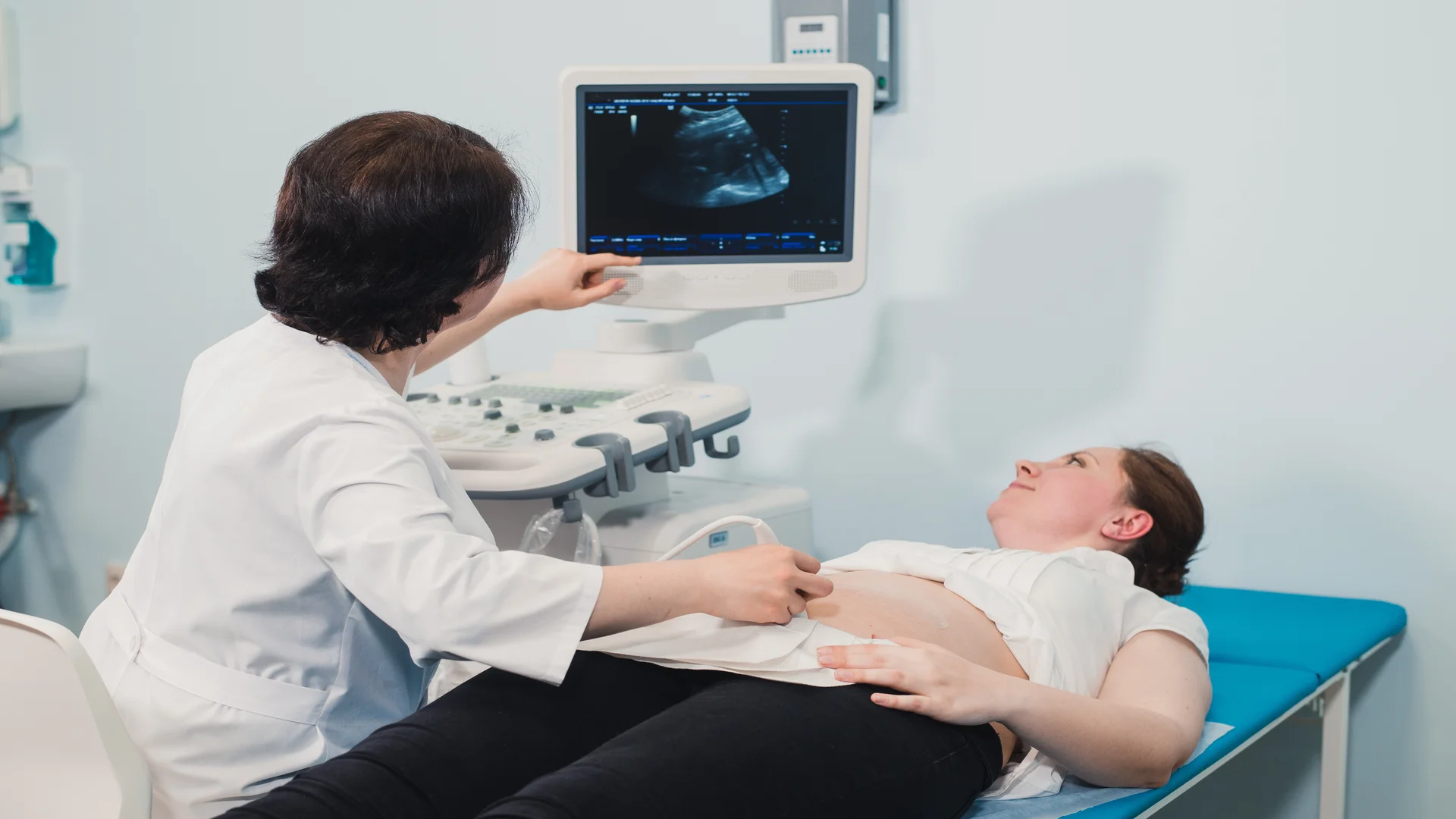
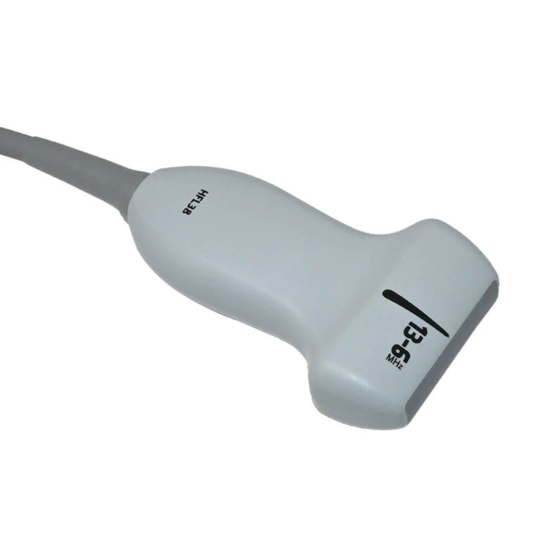
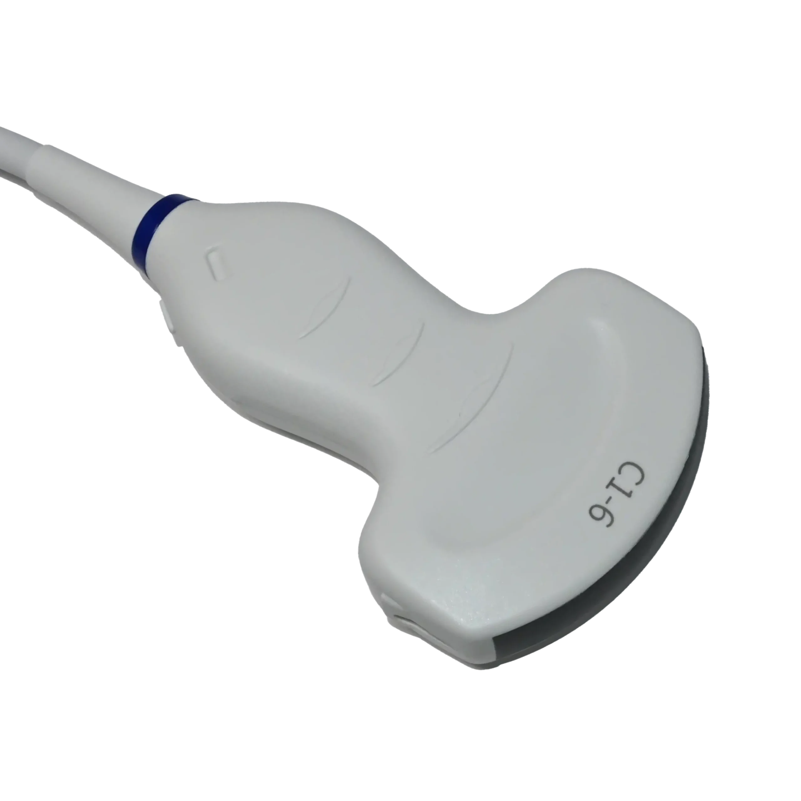
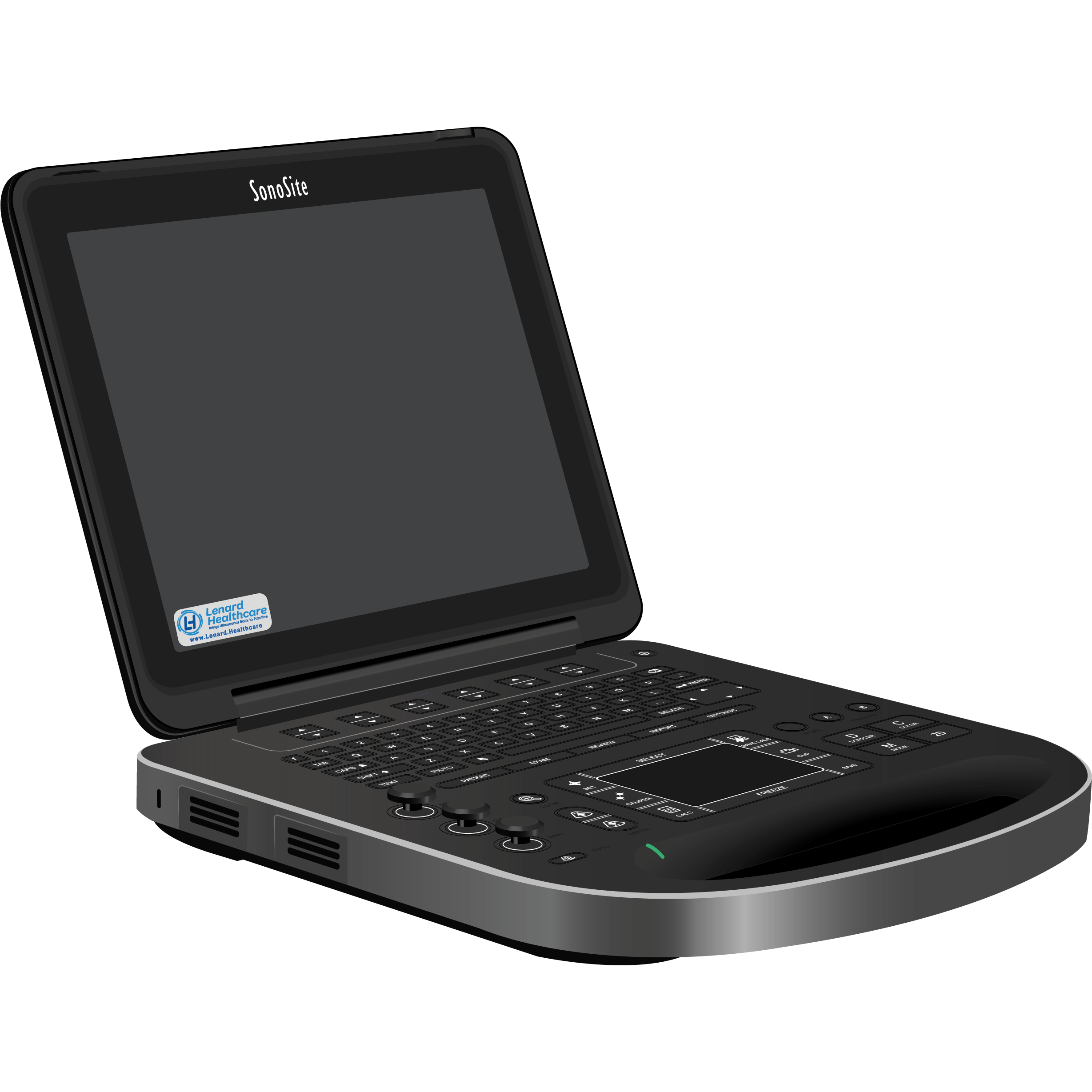
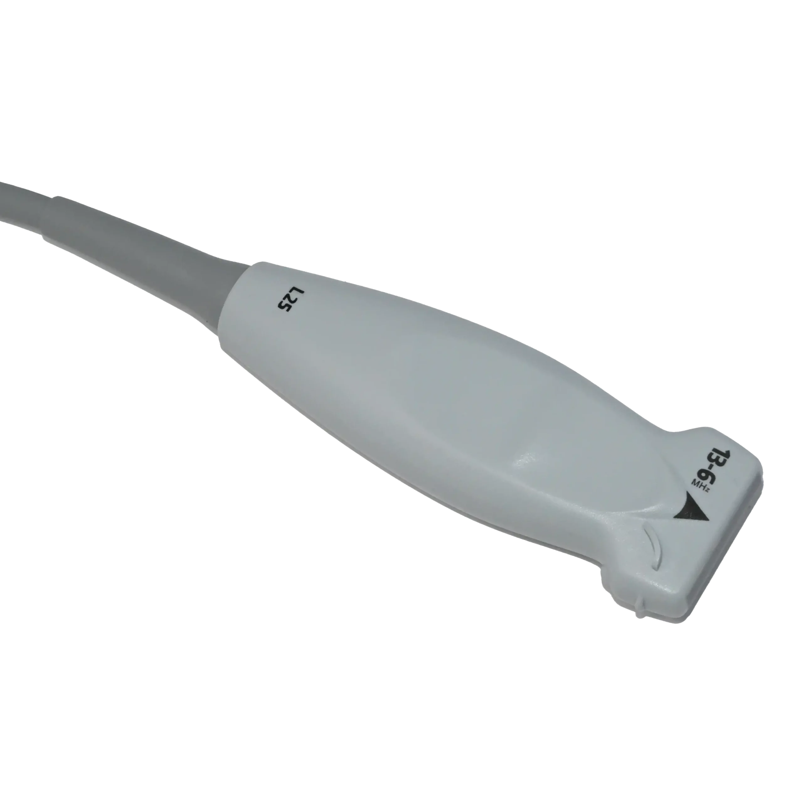
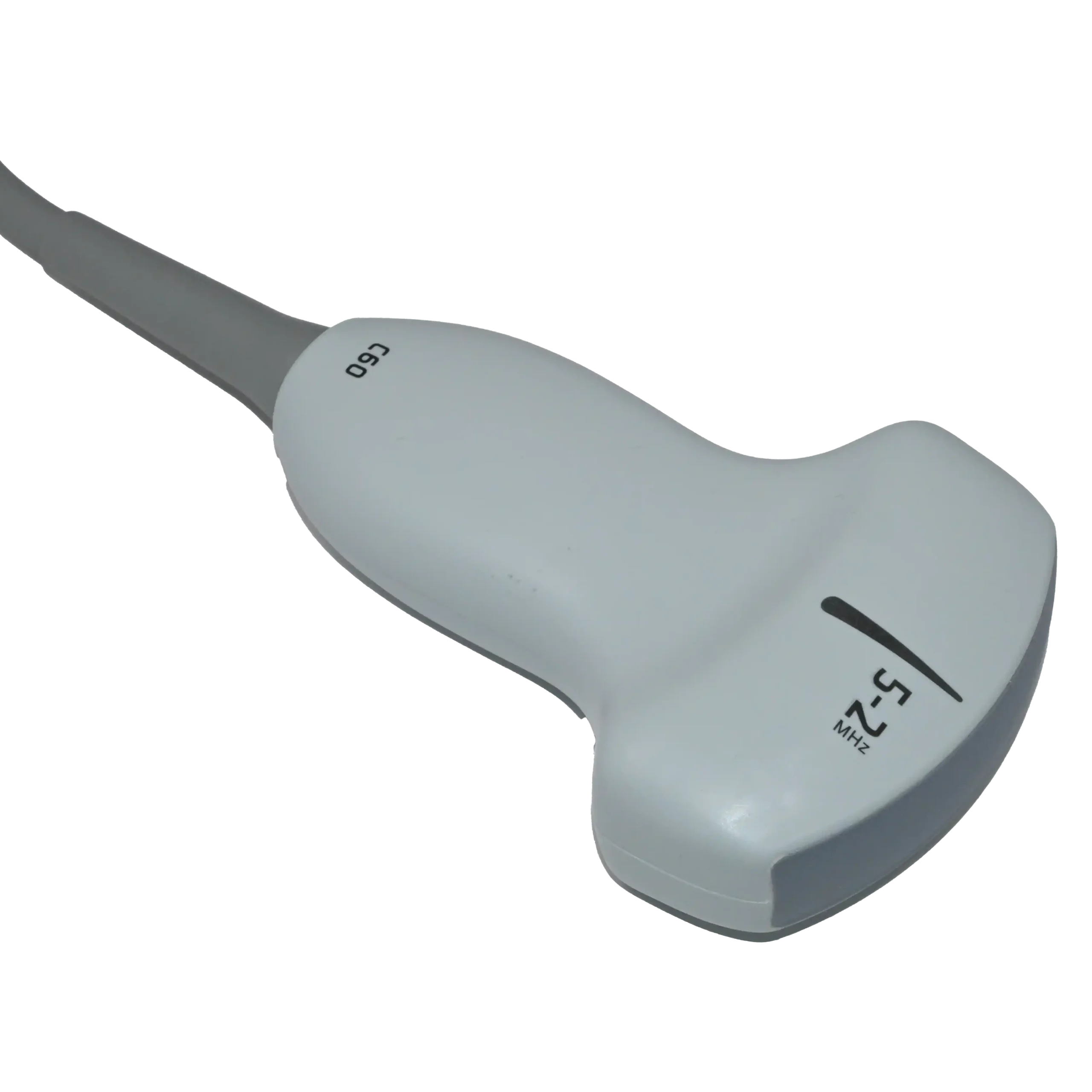
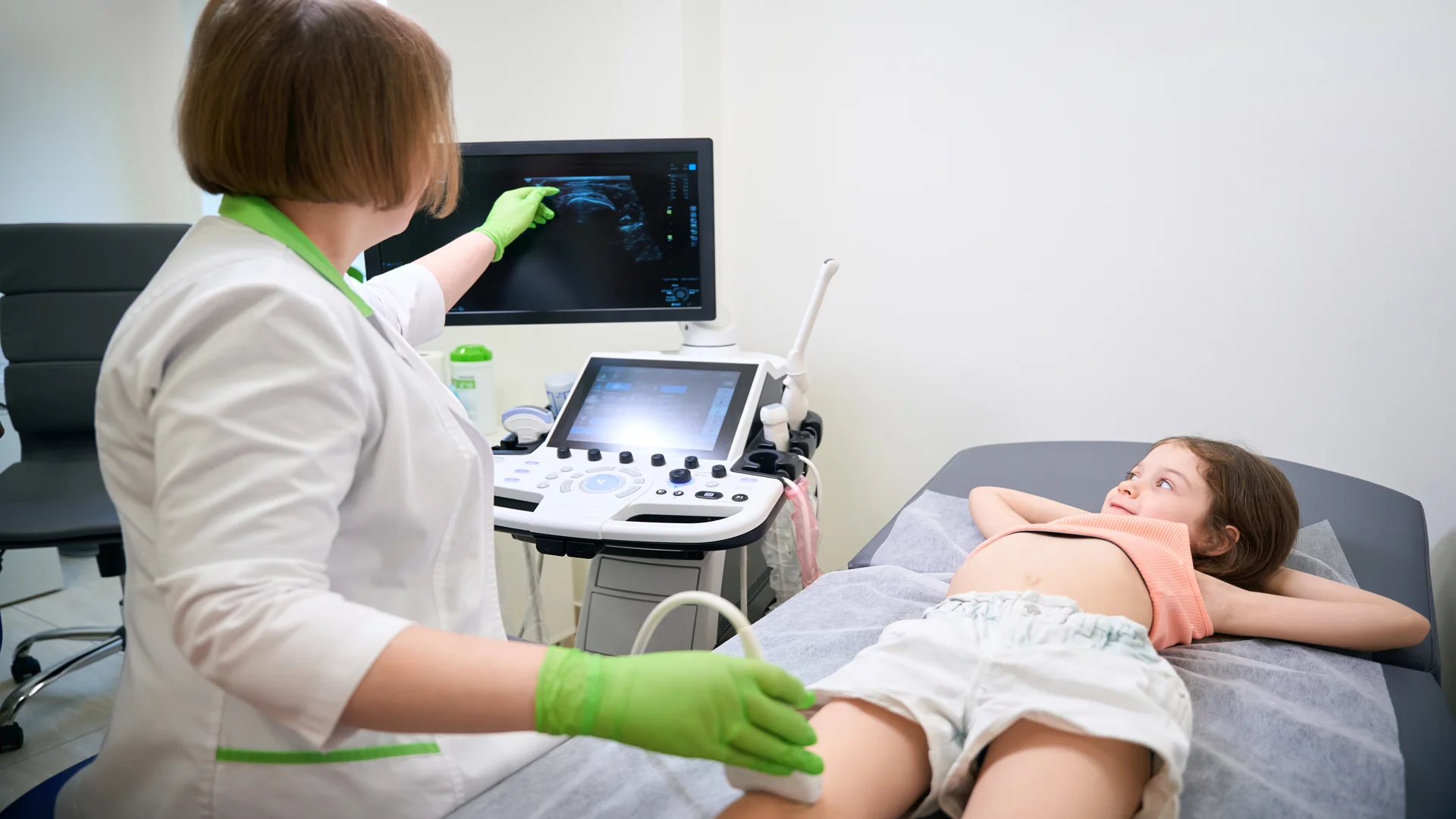
Among the different types of medical imaging, ultrasound is considered one of the safest methods because it does not use ionizing radiation. Instead, doctors use ultrasound machines equipped with different types of transducers to produce real-time images of organs, soft tissues, and blood flow. The choice of ultrasound transducer depends on the body area being examined and the required imaging depth.
Common ultrasound techniques include:
- Abdominal Ultrasound: Examines internal organs such as the liver, gallbladder, pancreas, spleen, and kidneys.
- Obstetric and Gynecological Ultrasound: Assesses fetal development during pregnancy and examines the uterus and ovaries.
- Echocardiography (Cardiac Ultrasound): Evaluates the heart’s chambers, valves, structure, and pumping function; different cardiac ultrasound probes are used depending on the examination approach.
- Musculoskeletal Ultrasound: Examines muscles, tendons, ligaments, and joints, including certain injuries and inflammatory conditions.
Ultrasound machines come in different types, each designed for specific applications and clinical needs. To learn more, explore our comprehensive guide below, where we explain everything from the basics to practical applications.
Different Types of Ultrasound Machines
2. X-ray Imaging (Radiography)
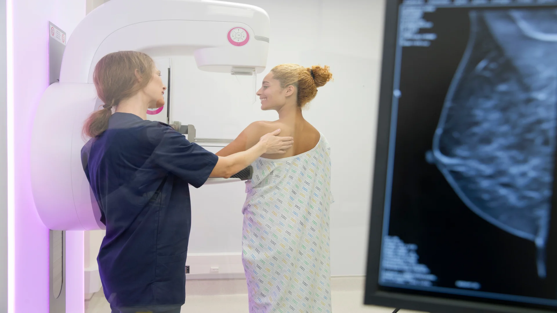
Conventional X-ray imaging passes a small amount of ionizing radiation through the body to create a projection image. Dense structures, such as bones, absorb more X-rays and appear lighter. It is commonly used to assess fractures, chest conditions, dental problems, and joint changes.
Common types of X-ray imaging include:
- Conventional Radiography: Produces static images, such as chest X-rays and bone examinations.
- Mammography: Uses low-dose X-rays to examine breast tissue and detect suspicious changes.
- Fluoroscopy: Produces moving X-ray images, often used in digestive examinations and image-guided procedures.
- Bone Density Scanning (DXA/DEXA): Measures bone mineral density to assess osteoporosis.
- Dental Radiography: Includes intraoral and panoramic X-rays used to examine teeth and surrounding structures.
3. Computed Tomography (CT) Scans
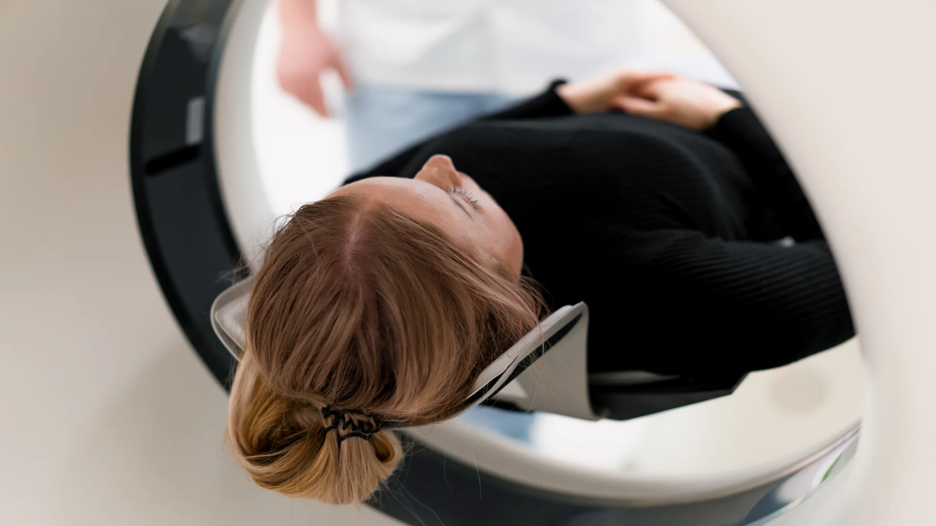
Computed tomography (CT) combines rotating X-ray equipment with computer processing to create cross-sectional images of the body. These images can be reconstructed in different planes, helping clinicians assess internal injuries, bleeding, lung and abdominal conditions, blood vessels, and tumors.
Common CT techniques and examinations include:
- Helical (Spiral) CT: Collects imaging data continuously as the patient moves through the scanner.
- Multidetector CT (MDCT): Uses multiple detector rows to capture detailed images efficiently.
- CT Angiography (CTA): Uses contrast material to visualize blood vessels, such as those supplying the brain or heart.
- Cardiac CT: Includes coronary calcium scoring and coronary CT angiography.
- Low-Dose CT: Uses reduced radiation settings for selected examinations, including lung cancer screening.
- Cone-Beam CT (CBCT): Produces 3D images, commonly used in dental and maxillofacial examinations.
4. Nuclear Medicine Imaging
Nuclear medicine imaging uses small amounts of radioactive materials, known as radiopharmaceuticals, to examine how organs and tissues function. It is used in selected cancer, heart, bone, thyroid, and kidney assessments, providing functional information that can complement anatomical imaging.
The main nuclear medicine imaging techniques include:
- Planar Scintigraphy: Produces two-dimensional images of radiotracer distribution, as in thyroid and bone scans.
- Single-Photon Emission Computed Tomography (SPECT): Creates three-dimensional images, commonly used in cardiac perfusion examinations.
- Positron Emission Tomography (PET): Uses radiotracers to evaluate metabolic and other biological activity, often in cancer assessment.
5. Magnetic Resonance Imaging (MRI)
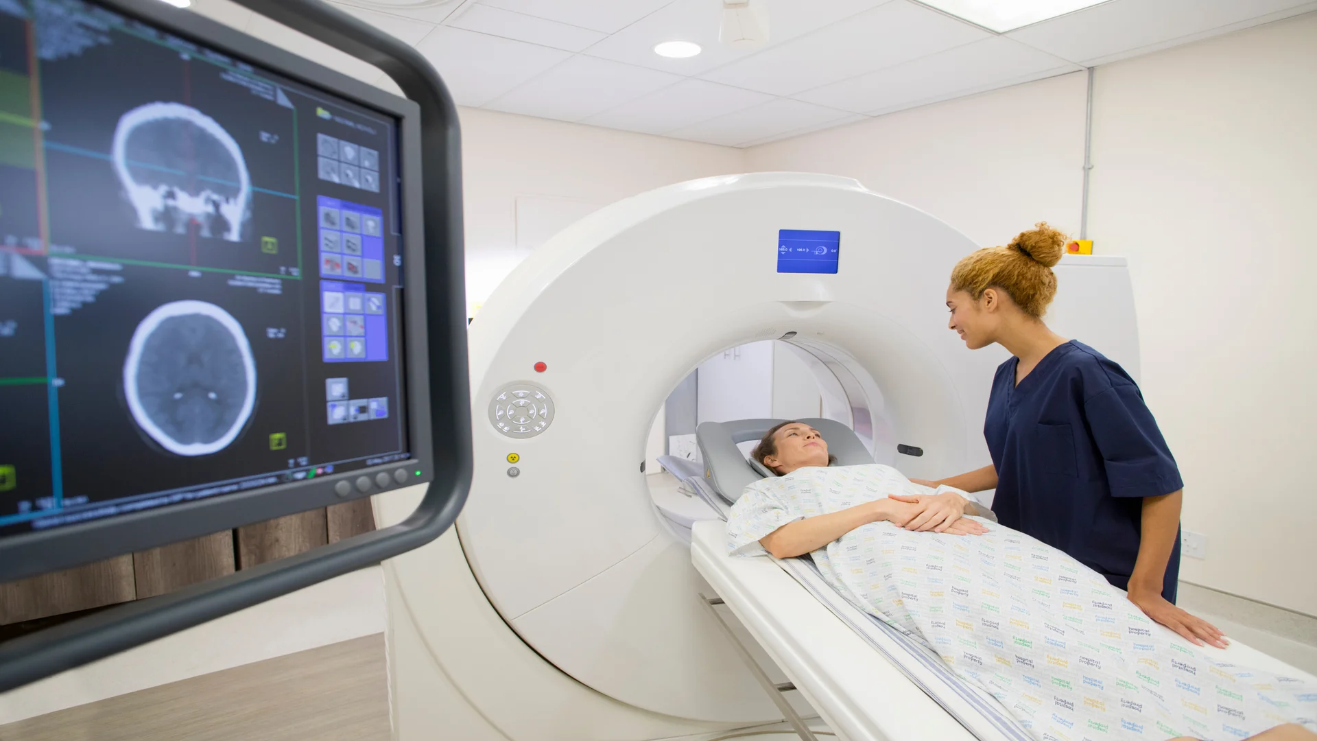
Magnetic resonance imaging (MRI) uses a strong magnetic field, radiofrequency pulses, and computer processing to create detailed images without ionizing radiation. It is particularly useful for examining the brain, spine, joints, muscles, and internal organs, including in the investigation of neurological conditions and soft-tissue injuries.
Common MRI techniques and examinations include:
- Functional MRI (fMRI): Maps changes in brain activity by measuring associated blood-oxygen changes.
- Diffusion-Weighted Imaging (DWI): Examines water movement within tissues and is particularly useful in acute stroke assessment.
- Magnetic Resonance Angiography (MRA): Produces images of blood vessels to investigate vascular conditions.
- Magnetic Resonance Cholangiopancreatography (MRCP): Examines the bile and pancreatic ducts.
- Magnetic Resonance Spectroscopy (MRS): Provides information about the chemical composition of selected tissues.
6. Hybrid Medical Imaging
Hybrid imaging combines two imaging technologies in one examination, pairing functional information with detailed anatomical images. It helps clinicians identify where abnormal activity occurs within the body and is commonly used in cancer assessment and selected cardiac and neurological investigations.
Common hybrid imaging systems include:
- PET/CT: Combines PET metabolic information with CT anatomical images.
- SPECT/CT: Combines SPECT functional information with CT anatomical detail.
- PET/MRI: Combines PET functional imaging with MRI’s detailed soft-tissue images.
How Do Doctors Choose Between Different Types of Medical Imaging?

Choosing among the different types of medical imaging depends on the suspected condition, the patient’s age, and the clinical information needed.
For example, when an infant has risk factors or examination findings suggesting hip dysplasia in babies, doctors may recommend a hip ultrasound to assess the development and position of the joint.
In a different clinical setting, a dentist investigating possible tooth decay may request targeted dental X-rays, such as bitewing images, to examine areas between the teeth that a visual examination alone cannot fully assess.
These examples illustrate how doctors select the most appropriate examination from the available types of medical imaging based on each patient’s diagnostic needs.
Comparing Different Types of Medical Imaging
Now that you have an overview of the different types of medical imaging, let’s bring them all together in the table below to make it easier to understand and compare the different types of medical scans.
| Imaging type | Technology | Uses ionizing radiation? | Common applications |
| Ultrasound | Sound waves | No | Pregnancy, heart, abdomen |
| X-Ray | X-rays | Yes | Bones, chest, teeth |
| CT | X-rays and computer reconstruction | Yes | Trauma, lungs, abdomen |
| MRI | Magnetic fields and radiofrequency signals | No | Brain, spine, joints |
| Nuclear medicine | Radiotracers | Yes | Cancer, heart, organ function |
Conclusion
When a physical examination alone isn’t enough to provide a clear diagnosis, different types of medical imaging help clinicians look deeper into the human body. From ultrasound and X-rays to MRI, CT scans, and other imaging techniques discussed in this article, each method offers unique insights to support diagnosis and treatment decisions.
But there’s more to explore! Stay tuned for our upcoming articles, where we’ll compare these types of medical scans in greater detail, highlighting their differences, advantages, limitations, and clinical applications.