When a patient walks in with shoulder pain, a swollen knee, or a suspected tendon injury, clinicians have more imaging options than ever, and choosing the right one matters. Musculoskeletal (MSK) ultrasound has become a go-to first step for many practices, offering real-time, radiation-free views of muscles, tendons, ligaments, and joints without the cost or wait times often associated with MRI.
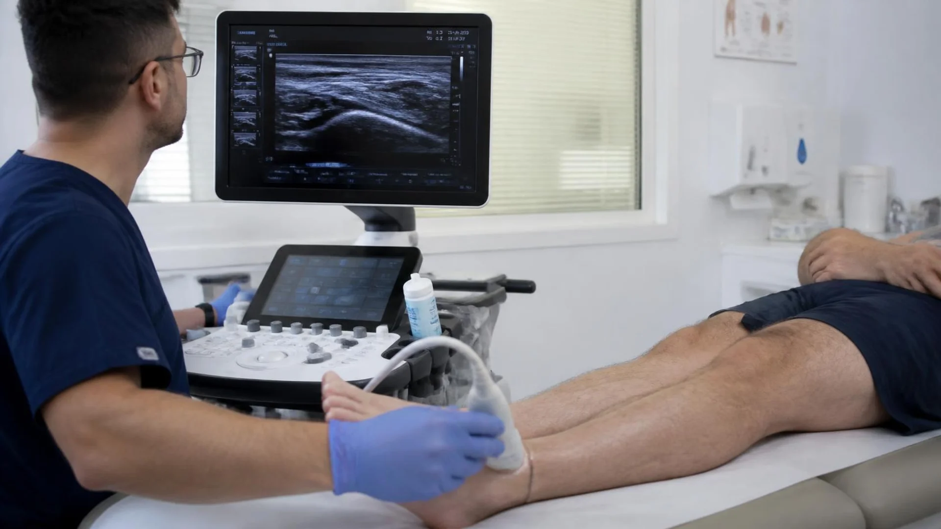
Unlike static imaging methods, MSK ultrasound allows clinicians to see structures as they move. They can watch an elbow bend, a foot flex, or a shoulder rotate to identify problems that may only appear during movement or under stress. It is also widely used to guide injections and aspirations, helping clinicians place the needle more accurately than landmark-based techniques.
In this guide, we’ll break down what musculoskeletal ultrasound is, how it compares to MRI and X-ray, where AI is starting to reshape the workflow in 2026, and what both clinicians and patients can expect from an exam, along with a realistic look at MSK price and equipment options.
What Is Musculoskeletal Ultrasound?
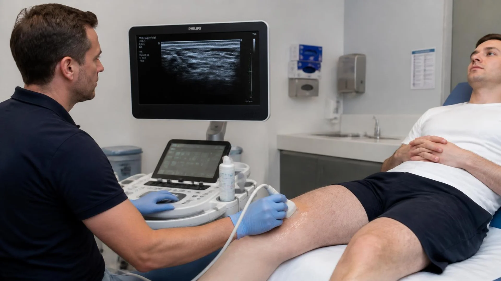
The term musculoskeletal refers to the body’s movement and support system, including the muscles, bones, joints, tendons, ligaments, and related soft tissues. Musculoskeletal ultrasound, also known as MSK ultrasound, is a medical imaging technique that generates real-time images of many of these structures. It allows healthcare professionals to examine soft tissues and joint-related structures without using ionizing radiation.
The technology behind MSK ultrasound follows the same fundamental principles as other forms of diagnostic ultrasound. Using an ultrasound system, whether a new or a properly maintained refurbished ultrasound machine, and an appropriate transducer (ultrasound probe), high-frequency sound waves are transmitted into the body. As these waves encounter tissues with different acoustic properties, some of their energy returns to the transducer as echoes. The ultrasound machine processes these returning signals to create images on the display in real time.
What Does Musculoskeletal Ultrasound Diagnose? (MSK Applications)
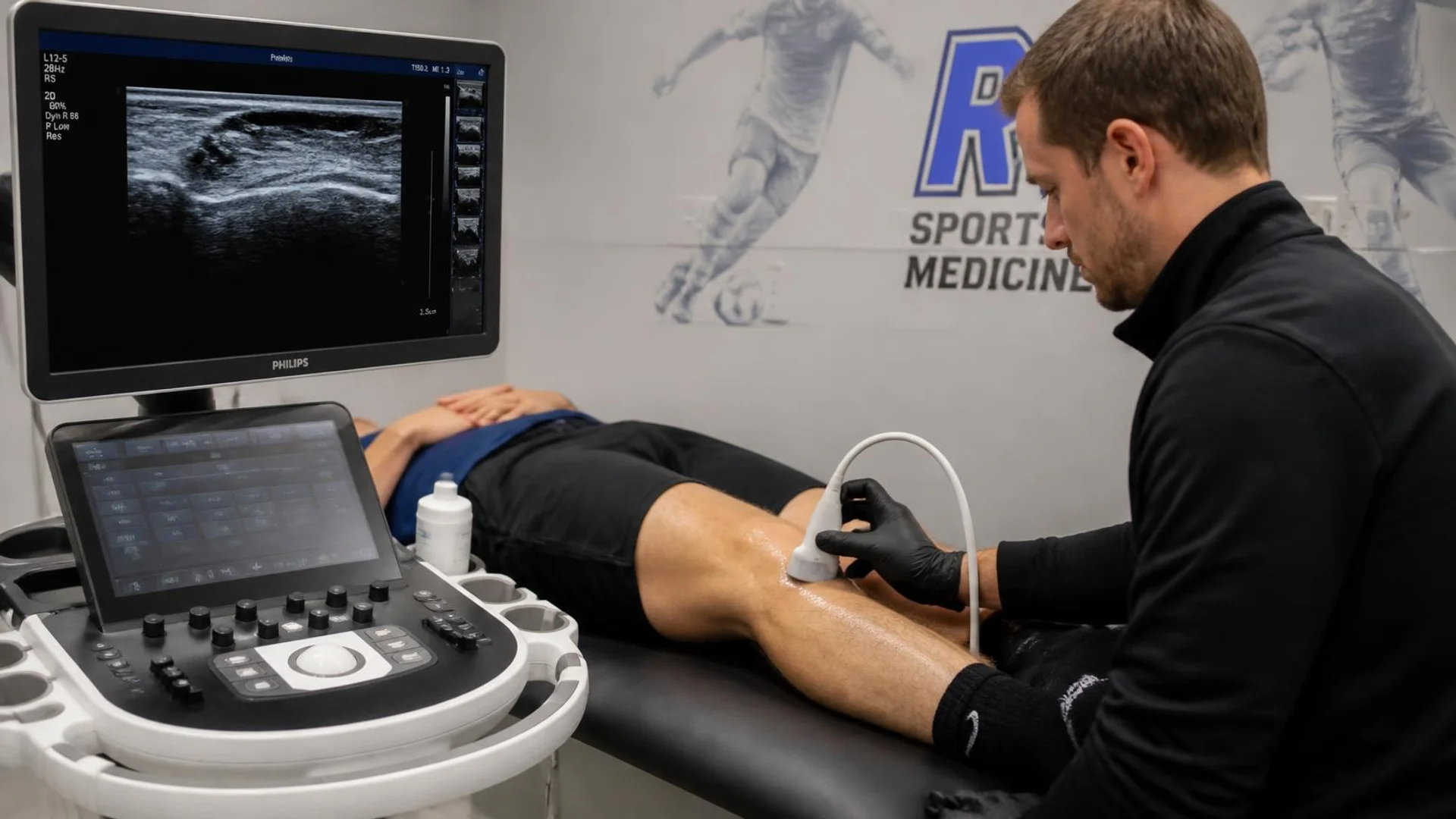
Musculoskeletal ultrasound can help diagnose a variety of conditions affecting tendons, ligaments, muscles, joints, bursae, and peripheral nerves. Common findings include tendon and muscle tears, tendinopathy, ligament injuries, bursitis, joint effusion, synovitis, and nerve entrapment. Depending on the clinical concern, MSK ultrasound can be performed around the shoulder, elbow, wrist, hand, hip, knee, ankle, and foot, as well as along individual muscles, tendons, and nerves.
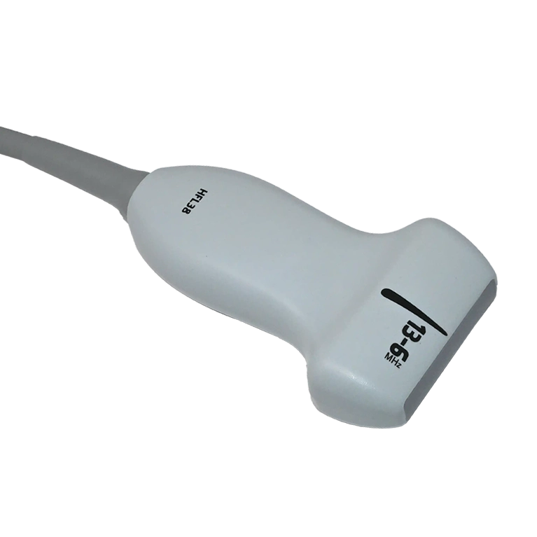
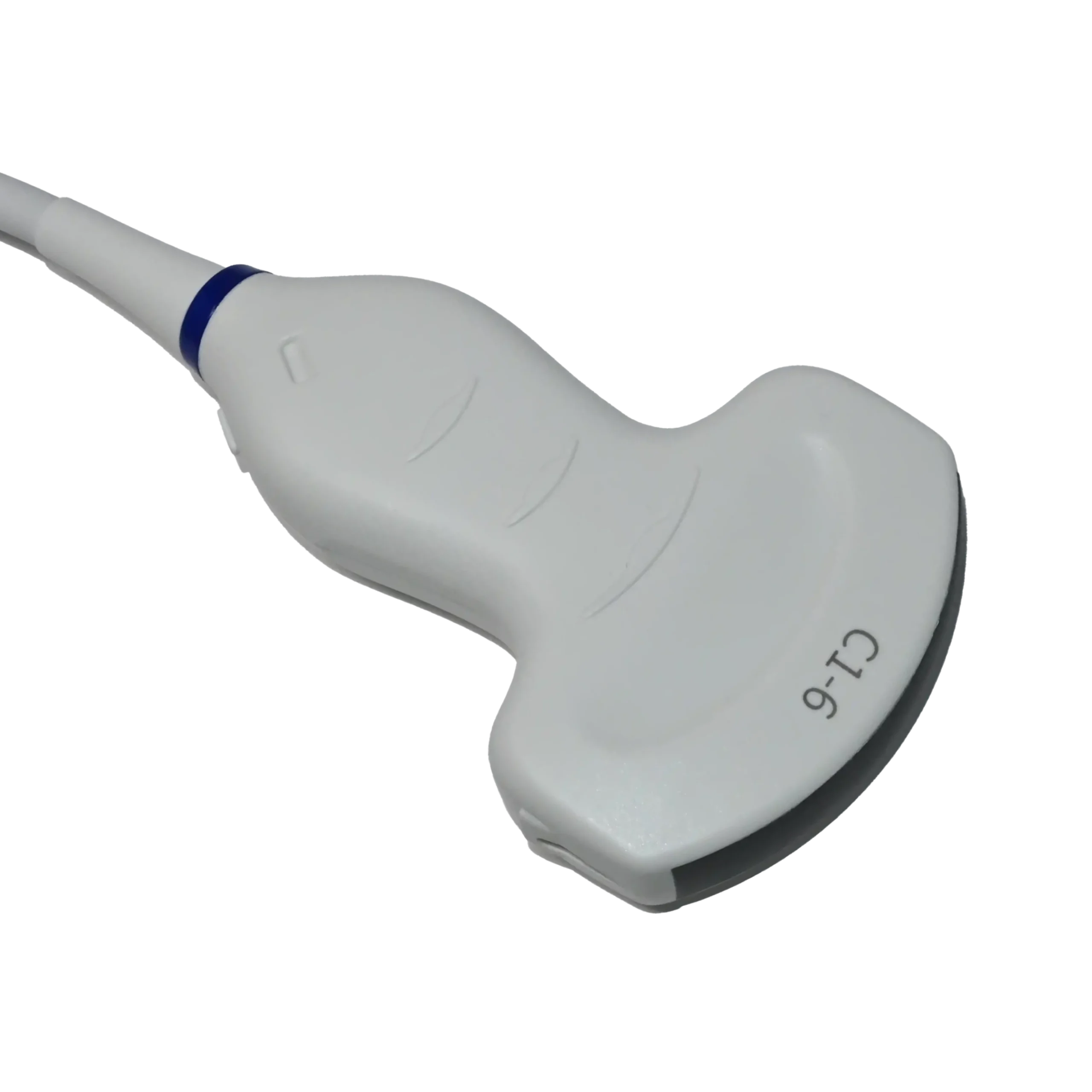
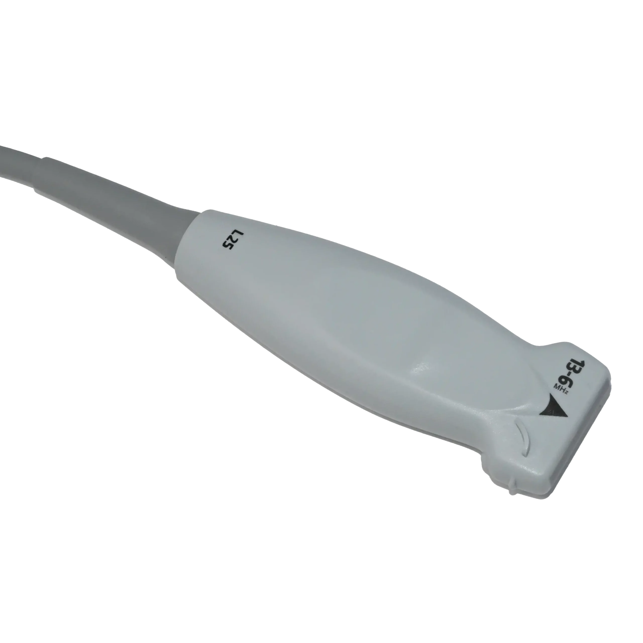
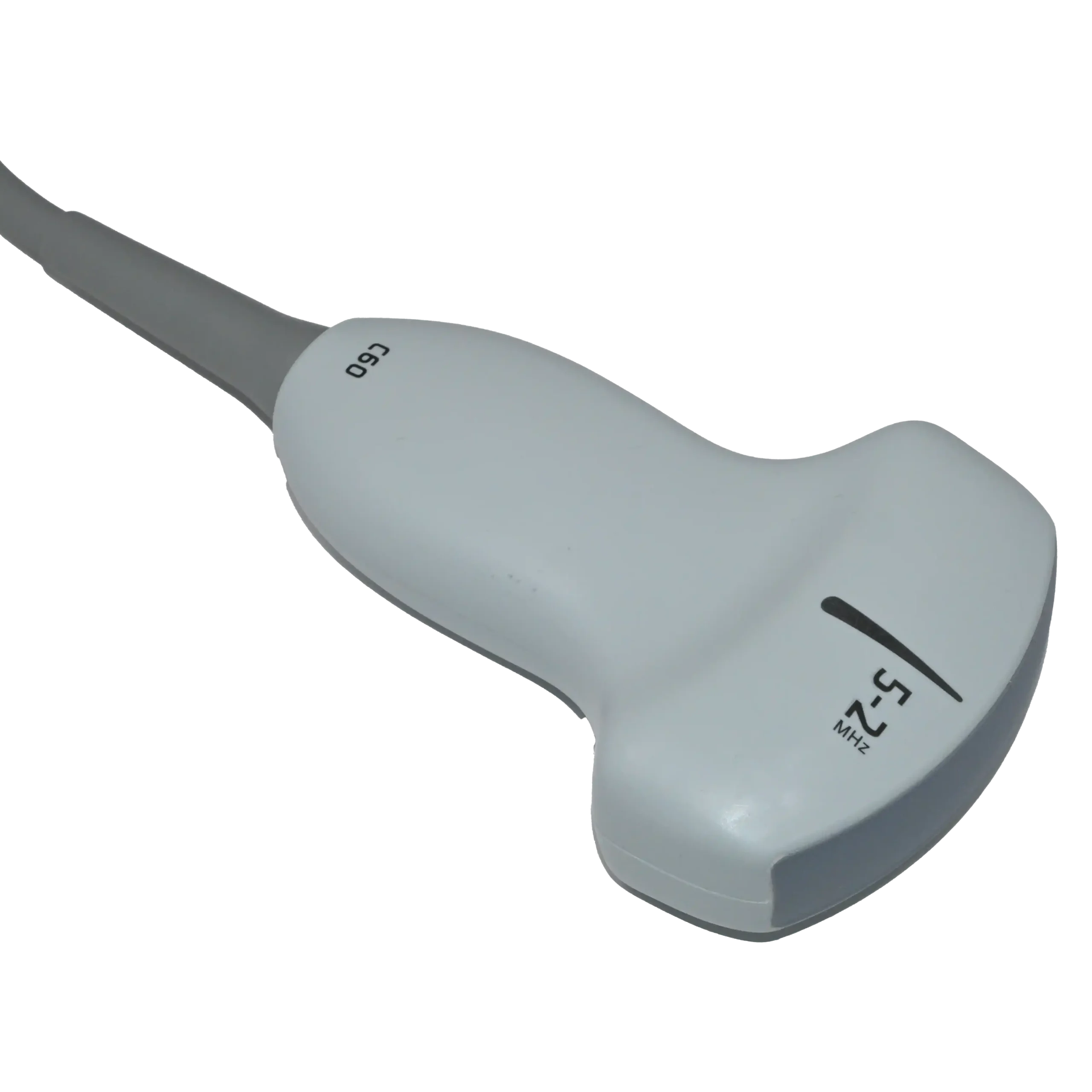
For example, after a football-related knee injury, a musculoskeletal ultrasound machine may be used to examine superficial structures such as the patellar or quadriceps tendon, medial collateral ligament, bursae, and joint fluid. The clinician selects the transducer according to the depth and location of the target. Generally, a high-frequency linear-array transducer is well suited to superficial tendons and ligaments, while deeper structures may require a lower-frequency linear or curvilinear probe for greater penetration.
Musculoskeletal Ultrasound vs MRI vs X-Ray
When a musculoskeletal problem needs imaging, ultrasound is not the only option. MRI and X-ray are also widely used, but each provides a different type of information and may be better suited to particular tissues or injuries. This often raises a practical question: when is MSK ultrasound used instead of MRI or X-ray, and what does each method show best?
MRI (Magnetic Resonance Imaging)
MRI uses magnetic fields and radiofrequency signals to create highly detailed images without ionizing radiation. It is particularly useful when clinicians need to examine deeper or more complex structures, including cartilage, bone marrow, menisci, ligaments, and other soft tissues.
X-ray
X-Ray imaging uses a small amount of ionizing radiation and is primarily used to examine bones. In musculoskeletal care, it is commonly requested when looking for fractures, changes in bone alignment, or features associated with conditions such as osteoarthritis.
MSK ultrasound
By comparison, MSK imaging is particularly useful for accessible soft tissues and allows structures to be examined dynamically while the patient moves. Depending on the anatomy and imaging depth, clinicians can select from different types of ultrasound transducers to obtain the required image quality.
| Factor | MSK Ultrasound | MRI | X-Ray |
| Best for | Tendons, ligaments, muscles, bursae, superficial nerves, joint effusion | Cartilage, bone marrow, menisci, deep ligaments, complex soft-tissue detail | Bone fractures, alignment, osteoarthritis, bone density changes |
| Radiation | None | None | Low-dose ionizing radiation |
| Imaging through bone | Limited; sound waves don’t penetrate bone well | Yes, can visualize structures adjacent to and within bone | Yes, designed specifically for bone |
| Real-time/dynamic imaging | Yes, structures can be assessed in motion | No, static images only | No, static images only |
| Field of view | Smaller, targeted area | Large, comprehensive | Large, comprehensive |
| Guides injections/procedures in real time | Yes | No | No |
| Typical exam time | 15–30 minutes | 30–60 minutes | Under 15 minutes |
| Relative cost | Lower | Highest | Lowest |
| Wait time/accessibility | Often available same-day, point-of-care | Usually requires scheduling; less portable | Widely available, minimal wait |
| Operator dependence | High, accuracy depends on examiner skill | Lower, standardized image acquisition | Lower, standardized image acquisition |
| Repeatable without added risk | Yes, no radiation exposure | Yes, no radiation exposure | Limited, cumulative radiation exposure |
Who Performs Musculoskeletal Ultrasound?
MSK ultrasound is used by a wide range of healthcare professionals, not just one specialty. Radiologists and sports medicine physicians are among the most frequent users, while rheumatologists, orthopedic surgeons, physiatrists, podiatrists, and chiropractors also incorporate it into their practice. Physical therapists are increasingly adopting MSK ultrasound as well, although their scope of practice varies by state and country.
With so many different providers using the technology, professional certifications play an important role in establishing consistent standards for training and competency. Two certifications are particularly recognized:
- RMSK, issued by APCA (Alliance for Physician Certification & Advancement), is built for physicians who perform and interpret MSK scans themselves.
- RMSKS, issued by ARDMS (American Registry for Diagnostic Medical Sonography), is designed for sonographers and technologists who perform scans on a physician’s behalf.
Put simply: RMSK is the physician’s credential; RMSKS is the sonographer’s. Both require passing a dedicated exam and meeting clinical eligibility requirements, and both are becoming a trusted signal of skill for patients and referring providers alike.
Ultrasound-Guided Injections & Procedures
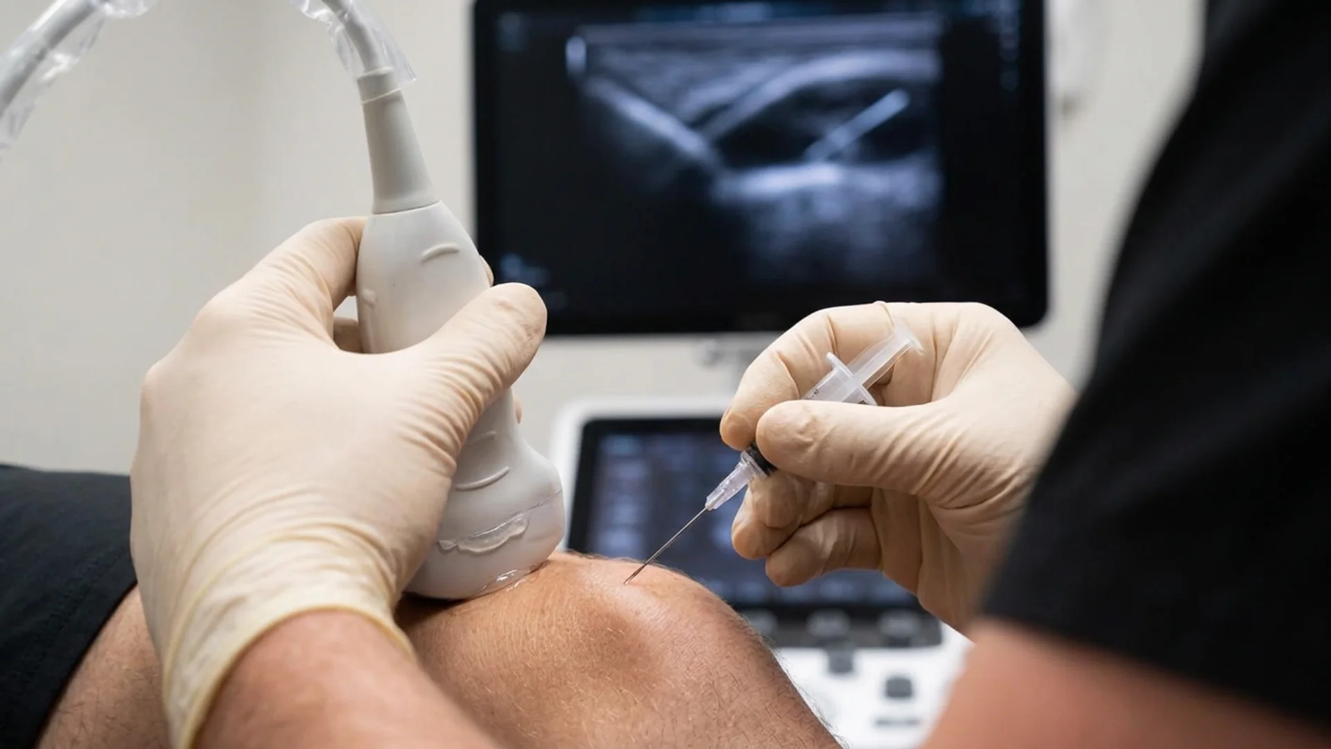
Musculoskeletal ultrasound is not limited to diagnosing injuries; it can also provide real-time guidance during injections and other minimally invasive procedures. Instead of relying only on anatomical landmarks, a technique sometimes called a “blind” or, more accurately, landmark-guided injection, the clinician can watch the needle advance on the ultrasound screen and guide it toward the intended joint, bursa, tendon sheath, or other target while avoiding nearby nerves and blood vessels. This direct visualization can improve the accuracy of needle placement for many musculoskeletal procedures.
A common example is an ultrasound-guided knee injection or aspiration. With an MSK ultrasound system and a suitable transducer, the clinician first identifies the joint space, surrounding tissues, and any fluid present, then follows the needle in real time as it approaches the target. For this type of procedure, a high-frequency linear probe designed for musculoskeletal imaging may be used, such as a refurbished Sonosite HFL38xi linear-array transducer, which is also compatible with an in-plane needle guide. The same real-time guidance can be applied to procedures such as joint aspiration, bursal and tendon-sheath injections, and selected perineural injections.
AI & Musculoskeletal Ultrasound in 2026
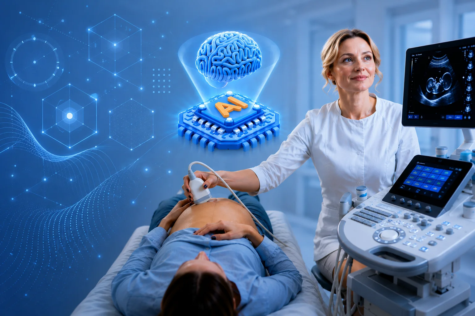
In 2026, artificial intelligence is becoming increasingly integrated into ultrasound technology. It helps clinicians with tasks such as image acquisition, optimization, measurements, and workflow automation. A good example is the refurbished Philips EPIQ Elite ultrasound, suitable for MSK applications, that incorporates AI-assisted and automated technologies to make imaging workflows more efficient and reproducible. Philips has continued to expand these AI capabilities through its 2026 Elevate Plus release, adding additional AI-powered workflow and measurement tools.
As AI continues to develop, its role is likely to remain supportive, helping imaging professionals work more efficiently while leaving clinical interpretation and decision-making in qualified hands. To learn more about the current applications and advances of this technology across healthcare, read our complete guide to AI in medical imaging.
Benefits of Musculoskeletal Ultrasound
So far, we have covered what MSK ultrasound is, where it is used, and the conditions it can help evaluate. Overall, the main benefits of musculoskeletal ultrasound include:
- Real-time imaging: Clinicians and healthcare providers can examine muscles, tendons, joints, and other structures as the patient moves.
- No ionizing radiation: MSK ultrasound is an imaging technique that uses sound waves rather than the radiation used in X-ray and CT imaging.
- Detailed soft-tissue imaging: Using high-frequency ultrasound probes can provide detailed images of many superficial tendons, ligaments, muscles, bursae, and peripheral nerves.
- Dynamic assessment: Structures can be evaluated during movement, helping to identify abnormalities that may not be apparent at rest.
- Guidance for procedures: Live imaging can aid clinicians in positioning needles during injections, aspirations, and other selected procedures.
- Fast and accessible: MSK ultrasound examinations can often be performed relatively quickly in clinics, imaging centers, and point-of-care settings.
- Portable imaging options: Compact, handheld ultrasound systems such as the new and refurbished Sonosite ultrasound machines can make MSK imaging more accessible at the point of care when appropriate.
- Repeatable examinations: Because ultrasound imaging does not use ionizing radiation, follow-up examinations can be performed when clinically necessary without adding radiation exposure.
Limitations of MSK Ultrasound
Although MSK ultrasound imaging offers numerous benefits, it also has several limitations that can affect when and how it is used:
- Operator dependence: Image quality and diagnostic accuracy depend considerably on the examiner’s training, technique, and experience.
- Limited imaging through bone: Ultrasound waves do not penetrate bone effectively, making structures inside or behind bone difficult to assess.
- Challenges with deeper structures: Image quality may decrease when examining anatomy located deeper within the body.
- Smaller field of view: Ultrasound typically evaluates a more targeted anatomical area than modalities such as MRI.
- Patient-related challenges: Body composition, swelling, limited mobility, or difficulty positioning the patient can sometimes make scanning more challenging.
- Not suitable for every condition: Certain bone, cartilage, bone-marrow, and deep joint abnormalities are generally better evaluated with MRI, X-ray, or CT.
- Potential imaging artifacts: Effects such as anisotropy can alter the appearance of tendons and other structures if the transducer angle is not properly maintained.
| Benefits | Limitations |
| Live, motion-based visualization | Skill-reliant results |
| Radiation-free technique | Poor penetration of bone |
| High-resolution soft-tissue views | Reduced clarity at depth |
| Safe for repeat scans | Narrower scanning area |
| Live-guided procedures | Positioning-sensitive scans |
| Quick, convenient scheduling | Not a fit for every condition |
| Compact, point-of-care systems | Angle-sensitive imaging |
What to Expect During a Musculoskeletal Ultrasound
As you now know, Musculoskeletal ultrasound is a non-invasive imaging method, with its main benefits and limitations discussed above. What to expect from the examination differs slightly depending on whether you are performing the scan or receiving it as a patient.
For the Clinician or Ultrasound Performer
A reliable MSK examination requires both appropriate equipment and a systematic scanning technique. Important considerations include:
- Review the patient’s history and clinical question.
- Position the patient appropriately for the target anatomy.
- Select the right transducer and frequency for the imaging depth.
- Optimize settings such as gain, depth, and focus.
- Examine relevant structures in appropriate imaging planes.
- Use dynamic assessment when clinically useful.
- Be aware of artifacts such as anisotropy.
For the Patient
Most diagnostic MSK ultrasound examinations require minimal preparation and can be completed without significant discomfort. Before and during the scan, you need to:
- Wear clothing that allows easy access to the area being examined.
- Remove jewelry or other items covering the scanning area if requested.
- Expect ultrasound gel to be applied to your skin.
- Be prepared to reposition or move the affected body part.
- Tell the examiner if pressure over an injured area becomes uncomfortable.
- Follow any additional preparation instructions provided by your healthcare professional.
How Much Does a Musculoskeletal Ultrasound Cost in 2026?
The price of MSK ultrasound equipment varies depending on the manufacturer, model, system configuration, transducers, imaging features, and equipment condition. New ultrasound machines generally come at a higher price, while used and refurbished systems can significantly reduce the initial investment. However, purchasing used equipment as-is may come with uncertainties regarding its condition, service history, and warranty coverage.
For facilities working within a defined budget, professionally refurbished MSK ultrasound systems can offer a practical balance between cost and reliability when they have been properly inspected and tested. Elzhen provides refurbished ultrasound equipment with a one-year warranty and after-sales support. Because pricing differs between models and configurations, you can request a quote and specify the system you are interested in to receive its current price and availability.
Conclusion
Musculoskeletal ultrasound has earned its place as a first-line imaging option for tendons, ligaments, muscles, and joints, thanks to its real-time, radiation-free views of structures in motion. That said, it isn’t meant to replace other imaging methods such as MRI or X-ray: deeper anatomy, bone, and certain cartilage findings still need those modalities for a complete picture.
Where ultrasound excels instead is speed, accessibility, and its ability to guide injections and aspirations with a precision that landmark-based techniques can’t match. As AI-assisted tools continue to streamline acquisition and measurement, MSK ultrasound is set to become an even more central part of everyday diagnostic and interventional care.