When talking about the different types of ultrasound machines, the word “type” can have different meanings. Depending on the context, it may refer to how a system is designed, what it is used for, the imaging capabilities it offers, or whether the machine is new, used, or refurbished.
Because there is no single way to categorize ultrasound equipment, this guide looks at ultrasound machines from each of these perspectives to give you a clearer picture of the options available.
What Are the Different Types of Ultrasound Machines?
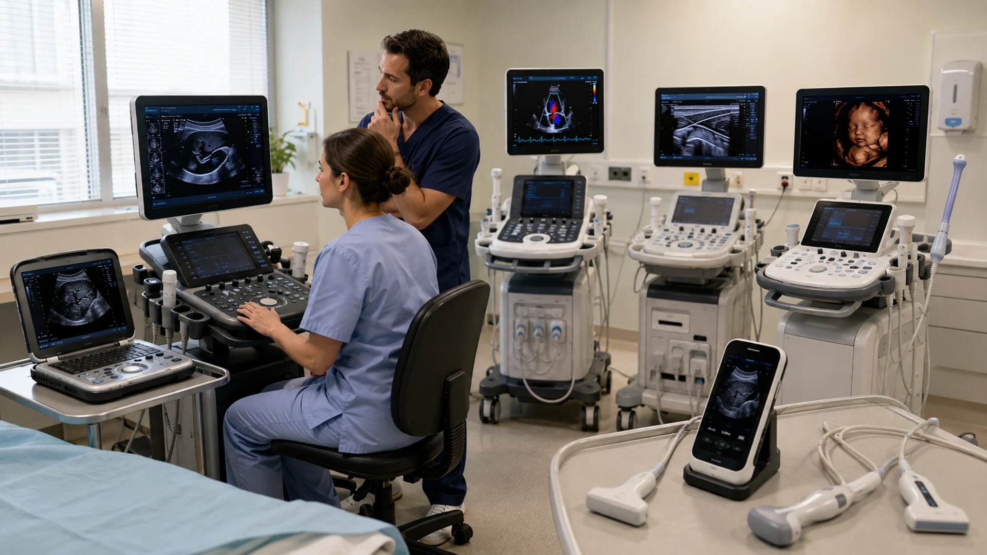
Ultrasound machines can generally be classified in several ways: physical design and portability, medical application, imaging modes and technologies, and system condition.
Physical design distinguishes systems such as console and portable units, while medical application groups them by areas such as cardiac, OB/GYN, vascular, and musculoskeletal imaging.
These systems can also differ in the imaging capabilities they support, from 2D ultrasounds to 3D/4D imaging and elastography. Finally, their condition separates them into new, used, and refurbished ultrasound machines.
Refurbished Ultrasound Machines
Let’s start with their physical design and take a closer look at the different types of ultrasound machines within this category.
Different Types of Ultrasound Machines by Physical Design
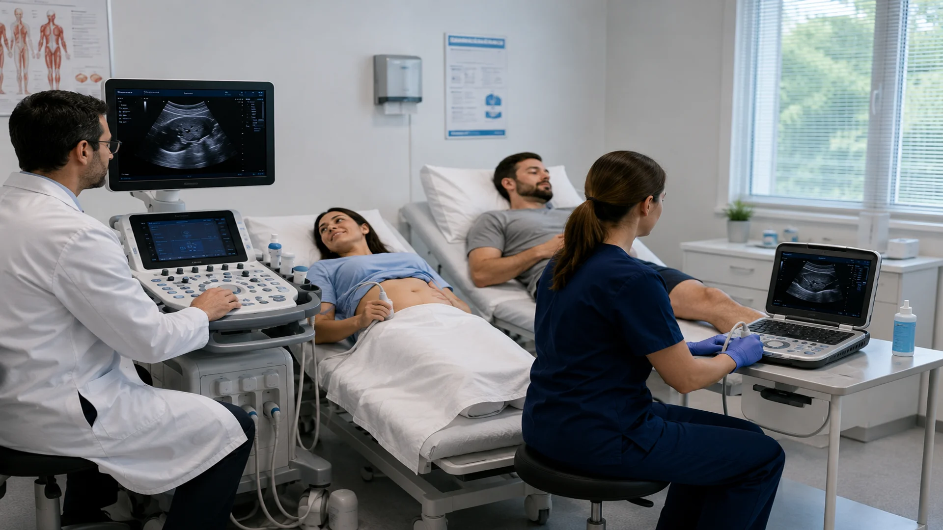
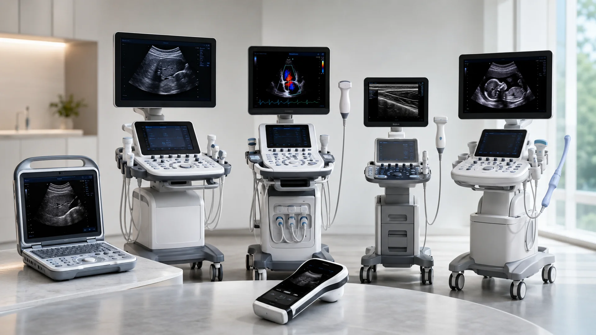
Based on their physical design and mobility, ultrasound machines can generally be divided into two main groups: console-based ultrasound and portable ultrasound machines. The main difference lies in how the system is built and moved between clinical environments, although their imaging capabilities can also vary considerably.
Console-Based Ultrasound Machines
Console-based ultrasound machines are full-size systems built around a wheeled workstation, typically with a larger display, integrated controls, and connections for multiple transducers. GE Healthcare is a major manufacturer in this category, offering console systems for applications such as general imaging, women’s health, and cardiovascular care.
For facilities looking for a lower-cost option than buying new, refurbished GE ultrasound machines are also available in a range of models and configurations.
Portable Ultrasound Machines
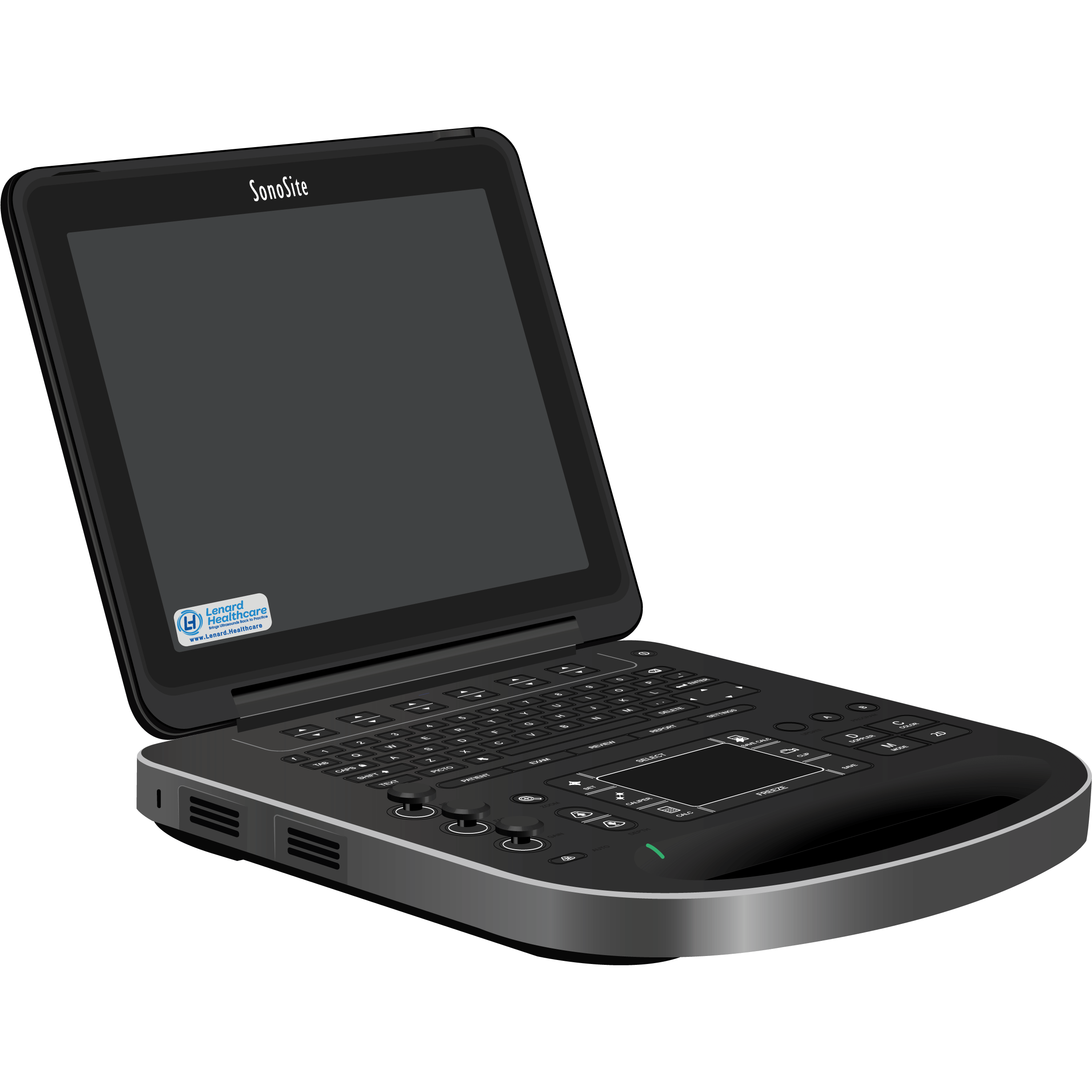
Portable ultrasound machines provide ultrasound imaging in a smaller, more mobile format, making them suitable for bedside examinations, point-of-care imaging, smaller clinics, and other settings where mobility matters.
Fujifilm Sonosite is particularly known for portable ultrasound systems, with compact models designed for use across a variety of clinical environments.
Types of Ultrasound Machines by Clinical Application
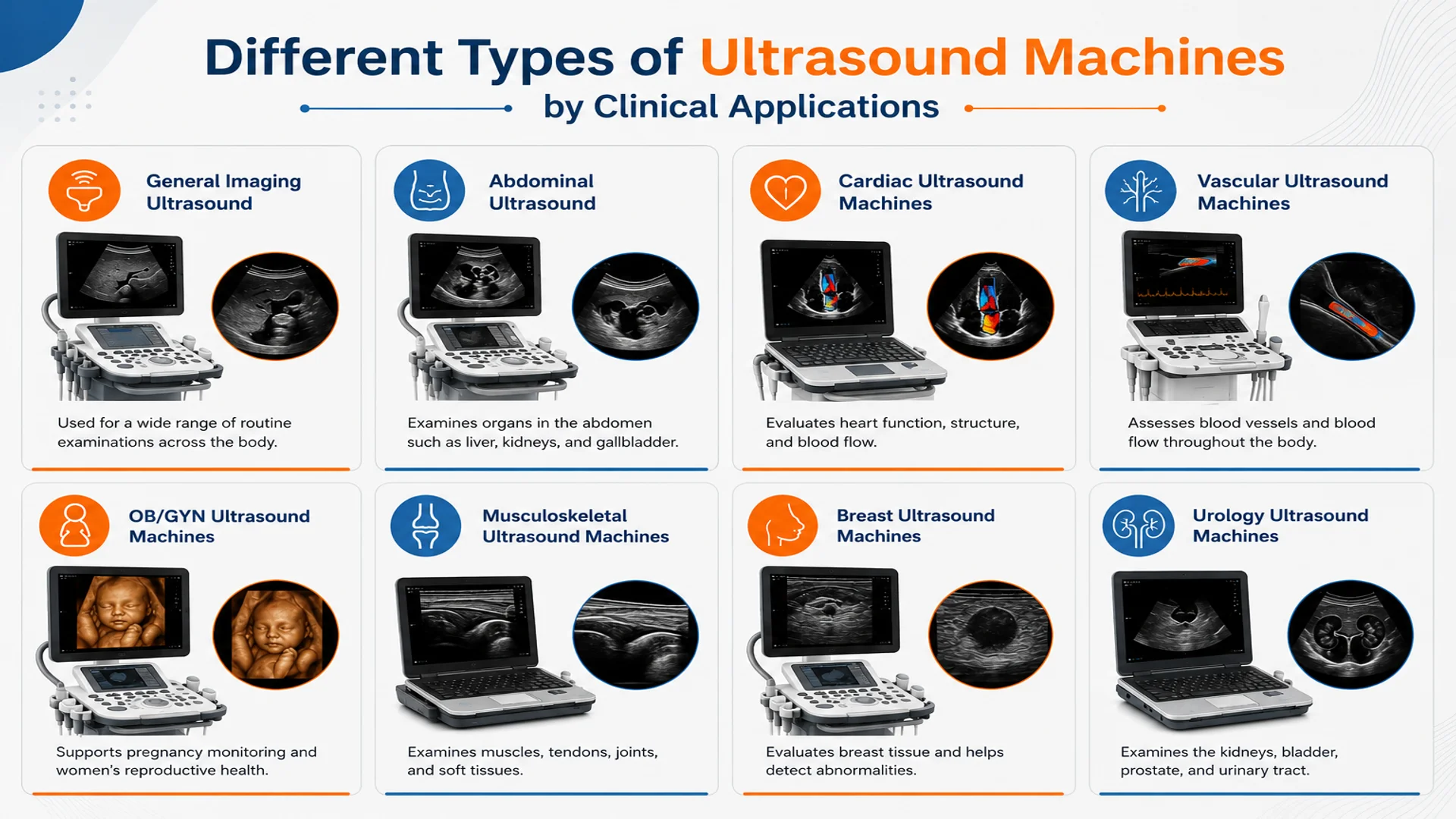
Another way to distinguish between different types of ultrasound machines is by their clinical application. The imaging needs of a cardiac examination, for example, are different from those of abdominal ultrasound imaging or an OB/GYN examination, and these differences can influence the system’s features, software, and compatible transducers.
For this reason, ultrasound systems are often configured for specific applications, including general imaging, abdominal ultrasound imaging, cardiology, OB/GYN, vascular imaging, musculoskeletal, and urology. Depending on the examination, different transducer types may also be used to provide the appropriate imaging depth, field of view, and level of detail.
General Imaging Ultrasound Machines
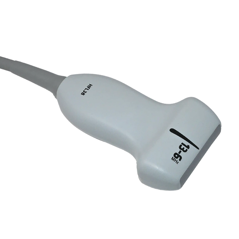
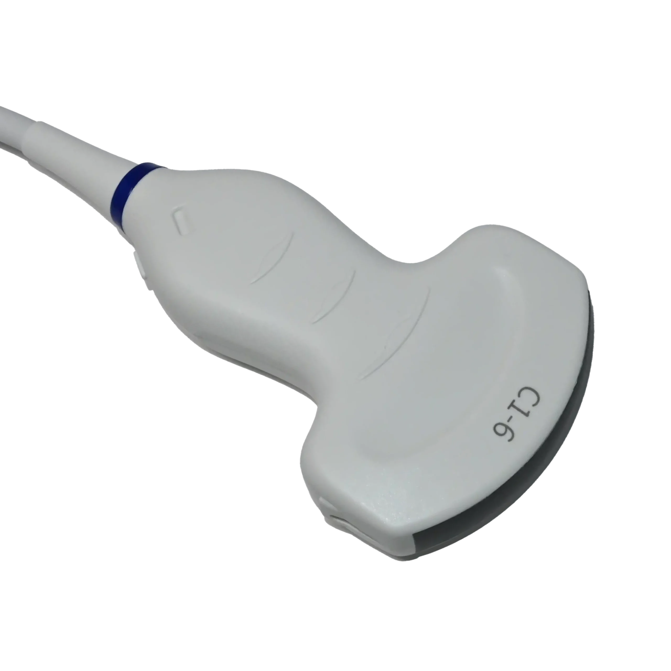
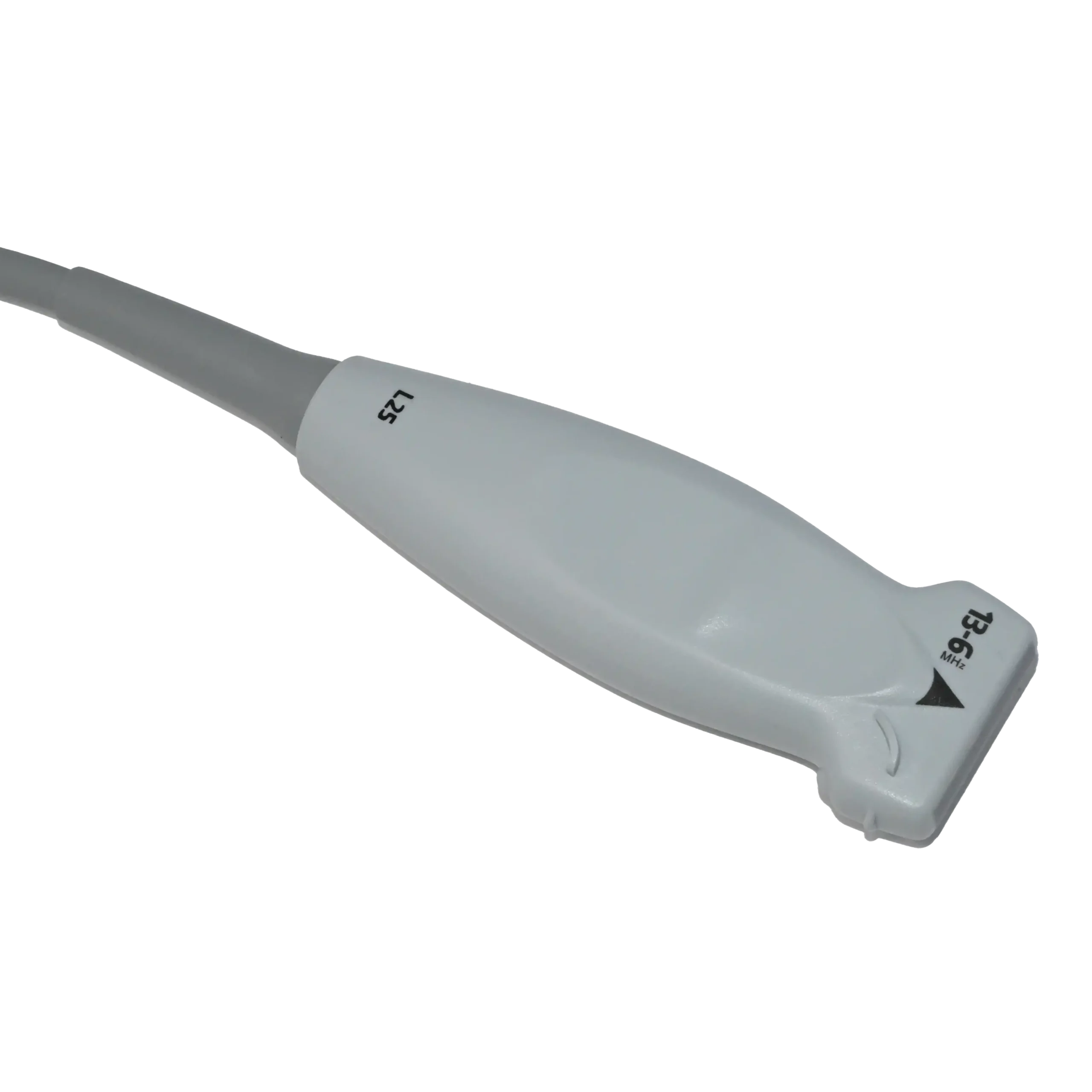
General imaging ultrasound machines, as their name suggests, are designed for a broad range of examinations rather than one specific specialty. They can be used for areas such as the abdomen, thyroid, breast, vascular system, and musculoskeletal structures, typically with convex probes for deeper abdominal imaging and linear probes for more superficial structures.
Abdominal Imaging Ultrasound Machines
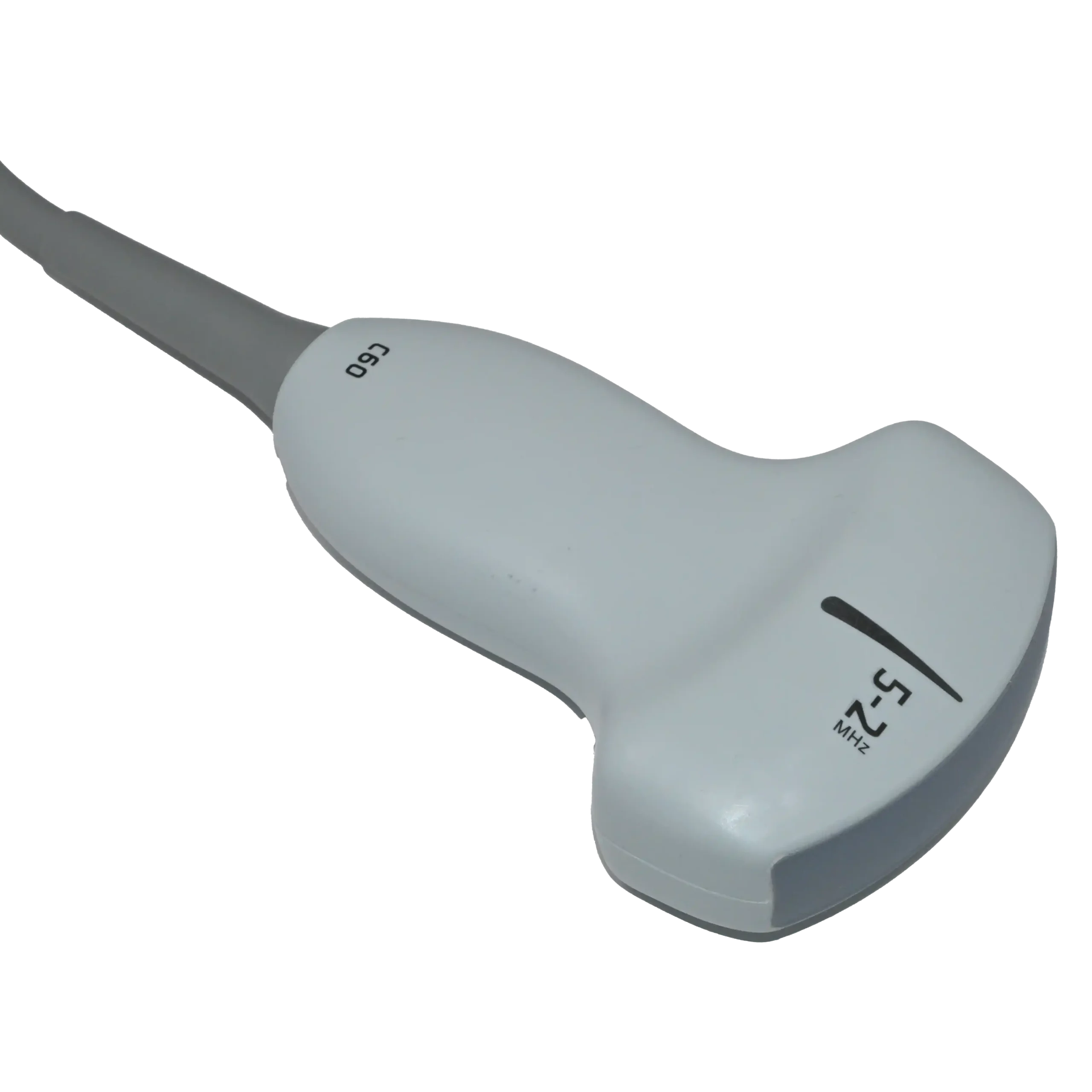
Abdominal ultrasound machines are used to examine organs and structures such as the liver, gallbladder, kidneys, pancreas, spleen, bladder, and abdominal blood vessels. A low-frequency convex (curvilinear) transducer, typically around 2–5 MHz, is commonly preferred because it provides the penetration and wide field of view needed for deeper abdominal structures.
Cardiac Ultrasound Machines
Cardiac ultrasound machines are configured for echocardiography, allowing clinicians to assess the heart’s structures, movement, and blood flow. They commonly use phased-array transducers, whose small footprint is well suited for imaging between the ribs, while Doppler modes help evaluate the direction and velocity of blood flow.
Vascular Ultrasound Machines
Vascular ultrasound machines are used to examine arteries and veins and evaluate how blood flows through them, making Doppler capabilities particularly important for these systems. Linear-array transducers are commonly used for peripheral vessels, while lower-frequency curved probes may be selected when deeper vessels need to be reached.
OB/GYN Ultrasound Machines
OB/GYN ultrasound machines are equipped for imaging throughout women’s healthcare, including pregnancy, fetal assessment, and gynecological examinations. Convex probes are commonly used for transabdominal scans, while transvaginal probes provide closer views of the uterus, ovaries, and early pregnancy; compatible volume probes can also provide 3D and 4D imaging.
Musculoskeletal Ultrasound Machines
Musculoskeletal (MSK) ultrasound machines visualize muscles, tendons, ligaments, joints, nerves, and surrounding soft tissues, including their movement during an examination. Because many of these structures are relatively superficial, clinicians commonly use high-frequency linear transducers to provide detailed imaging.
Breast Ultrasound Machines
Breast ultrasound machines are used to examine breast tissue and provide additional information about areas that may require further evaluation. Similar to the previous section, because breast structures are relatively superficial, these examinations typically rely on high-frequency linear-array transducers to produce detailed images.
Urology Ultrasound Machines
In urology, ultrasound provides real-time imaging of structures such as the kidneys, bladder, and prostate, supporting a range of urinary and male reproductive examinations. Convex probes are often suitable for viewing the kidneys and bladder through the abdomen, whereas transrectal transducers provide closer access to the prostate when more detailed imaging is required.
Types of Ultrasound Machines by Condition
Ultrasound machines can also be categorized according to their condition, particularly when comparing equipment available for purchase. The three common types of ultrasound machines by their condition are new, used, and refurbished systems, each representing a different equipment history and level of preparation before sale.
- New ultrasound machines: Systems sold without previous clinical use, typically supplied directly by the manufacturer.
- Used ultrasound machines: Previously owned or operated machines that are resold in their existing condition.
- Refurbished ultrasound machines: Previously used systems that have undergone inspection, testing, servicing, and, where necessary, repair or component replacement before being offered for sale again.
Types of Ultrasound Machines by Imaging Modes
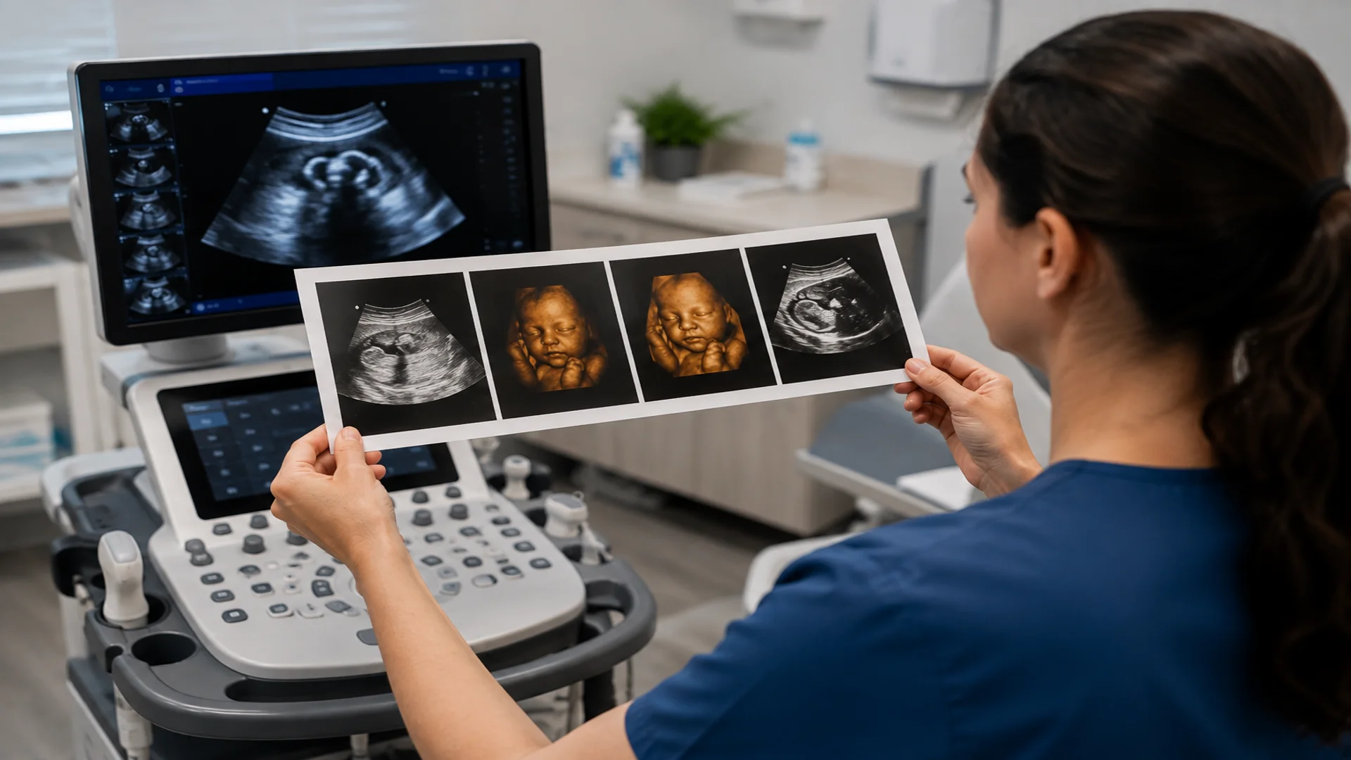
When exploring different types of ultrasound, you may come across terms such as 2D, Doppler, 3D, and 4D ultrasound, which are often described as ultrasound types. Although these imaging modes are not exactly separate types of ultrasound machines, and a single system can support several of them, they are still an important part of understanding how ultrasound systems differ.
So, to make the classification as clear and complete as possible, let’s take a closer look at the main ultrasound imaging modes and technologies and what each one offers.
B-Mode Ultrasound (2D Ultrasound)
B-mode gets its name from Brightness Mode and produces the familiar black, white, and gray ultrasound image seen on the monitor. The machine turns returning sound echoes into points of different brightness: stronger echoes appear brighter, weaker echoes appear in shades of gray, and fluid-filled areas often look black. Together, these points form a real-time 2D view of the anatomy.
For example, during an abdominal scan, B-mode can show the liver, kidneys, and gallbladder, allowing the clinician to examine their shape, position, and internal appearance.
M-Mode Ultrasound (Motion Mode)
M-mode stands for Motion Mode and shows how a structure moves over time. Instead of generating a full 2D view of an organ like B-mode, it follows a single ultrasound line and displays the movement along that line continuously across the screen. This makes it especially useful for assessing fast-moving structures, such as heart walls and valves, or for fetal heart motion.
Doppler Ultrasound
While B-mode and M-mode show anatomical structures and their movement over time, respectively, the Doppler imaging mode adds another layer of information by evaluating motion, particularly blood flow. It detects changes in the sound waves returning from moving blood cells, helping clinicians determine whether blood is flowing, its direction, and how fast it is moving.
Common Doppler modes and techniques used in diagnostic ultrasound include:
- Color Doppler: Adds color to the 2D image to represent blood flow and its direction.
- Pulsed-Wave (PW) Doppler: Measures blood-flow velocity at a specific location selected by the examiner.
- Continuous-Wave (CW) Doppler: Measures higher blood-flow velocities and is particularly useful in cardiac imaging.
- Power Doppler: Is sensitive to the presence of blood flow, including slower flow, but does not provide the same directional and velocity information as conventional Color Doppler.
More specialized Doppler techniques are also available. Tissue Doppler Imaging (TDI), for example, measures tissue movement rather than primarily focusing on blood flow and is commonly used in cardiac assessment. Manufacturers may also offer additional Doppler technologies developed for specific imaging applications.
3D Ultrasound
Moving beyond the two-dimensional images produced by conventional B-mode, 3D ultrasound creates a three-dimensional representation of the area being examined. The system collects ultrasound data from multiple planes and combines it into a volume that can be viewed and examined from different angles on the monitor.
A familiar example is 3D fetal ultrasound, which can provide a more recognizable view of features such as the fetus’s face and limbs. However, 3D imaging is not limited to pregnancy and can also be used in other areas, including gynecology and cardiac imaging.
4D Ultrasound
4D ultrasound is another term you may hear in clinical settings or come across when exploring different types of ultrasound machines. It takes imaging one step further than 3D ultrasound and, as its name suggests, adds a fourth dimension: time. This means that instead of displaying a still three-dimensional volume, 4D ultrasound shows that volume moving continuously in real time.
For example, during pregnancy, 4D ultrasound can allow the examiner to evaluate fetal movements, such as moving the hands, changing position, or making facial movements, as they happen on the monitor. The technology can also be used in other organs, including cardiac imaging, where real-time visualization of moving structures can be valuable.
Elastograph Ultrasound
Another term you may come across when exploring different types of ultrasound is ultrasound elastography. Rather than being a completely separate type of ultrasound system, elastography is a specialized imaging technology available on many ultrasound machines that provides information about tissue stiffness.
Two common approaches are strain elastography, which evaluates tissue deformation in response to compression or other mechanical forces, and shear-wave elastography, which measures how shear waves travel through tissue to assess stiffness. Elastography is used in several clinical areas, including liver, breast, thyroid, prostate, and musculoskeletal imaging.
Contrast-Enhanced Ultrasound (CEUS)
Last but not least, contrast-enhanced ultrasound (CEUS) is another term you may see when searching for different types of ultrasound machines, so it is worth briefly explaining this technology here as well.
CEUS uses a special contrast agent containing tiny gas-filled microbubbles that is injected into the bloodstream. These microbubbles make blood flow and tissue perfusion easier to see in real time, helping clinicians evaluate blood supply and characterize certain lesions, particularly in the liver.
B-mode, M-mode, Doppler, 3D/4D, elastography, and CEUS cover many common ultrasound modes and technologies, but the list does not end there. Modern systems may also include tissue harmonic imaging, panoramic or extended-field-of-view imaging, fusion imaging, and manufacturer-specific technologies, depending on the machine and its configuration.
Conclusion
As we have seen throughout this guide, different types of ultrasound machines can be distinguished in several ways, from their physical design and clinical applications to the transducers they support, their imaging technologies, and whether they are new, used, or refurbished.
With these classifications in mind, you should now have a clearer picture of the ultrasound systems you may encounter and how they differ.
Keep in mind that these categories are not always separate; a portable ultrasound machine, for example, may support several clinical applications, work with multiple transducers, and offer a range of imaging modes within a single system.