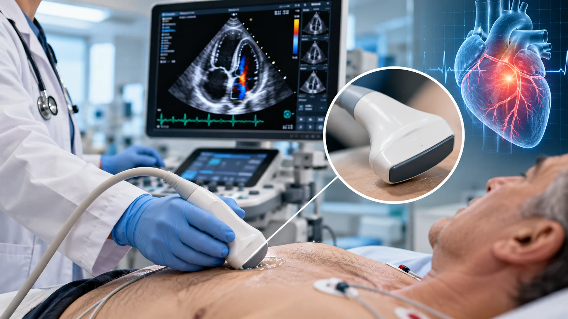

Some hip problems that affect people later in life can stem from conditions that begin in infancy. Developmental dysplasia of the hip (DDH) is one such condition that affects how a baby’s hip joint develops. Because signs are not always obvious, early assessment matters. Read on to learn about hip dysplasia in babies, how it’s diagnosed, and how pediatric hip ultrasound helps with diagnosis.
What Is Hip Dysplasia in Babies?
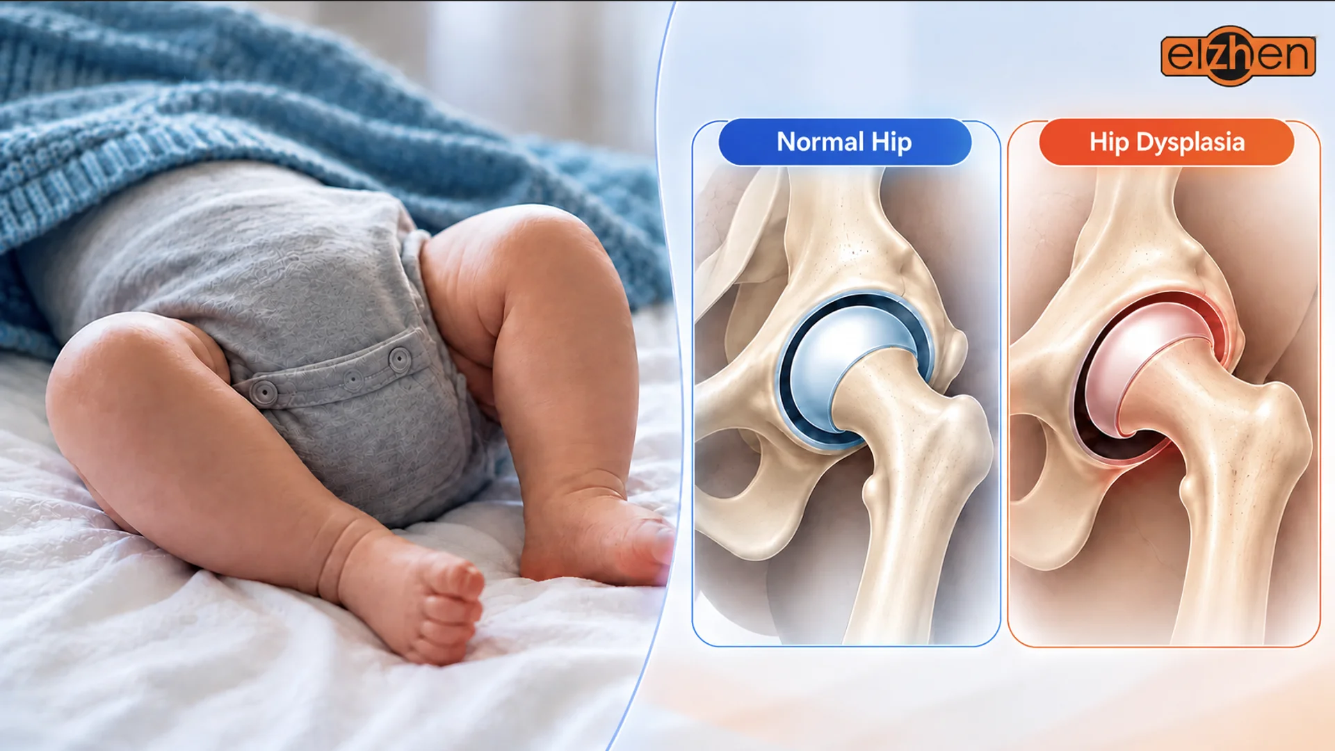
Hip dysplasia in babies, medically known as developmental dysplasia of the hip (DDH), is a condition in which the hip joint does not develop normally. In a healthy hip, the rounded head of the thighbone (femur) fits into a socket in the pelvis. With DDH, the socket may be too shallow to support the femoral head properly, or the joint may be unstable, allowing the bone to move partially or completely out of its normal position.
DDH is commonly identified during infancy, although some cases are diagnosed later in childhood. The condition may be present at birth or become apparent as the hip develops. Because affected babies may not show obvious signs, regular hip examinations are important, and clinicians may recommend an infant hip ultrasound when further assessment is indicated. This examination uses a suitable pediatric ultrasound probe to help clinicians evaluate the developing hip joint. In the following sections, we’ll explore how this imaging technique helps clinicians evaluate the developing hip joint.
Signs and Symptoms of Hip Dysplasia in Babies
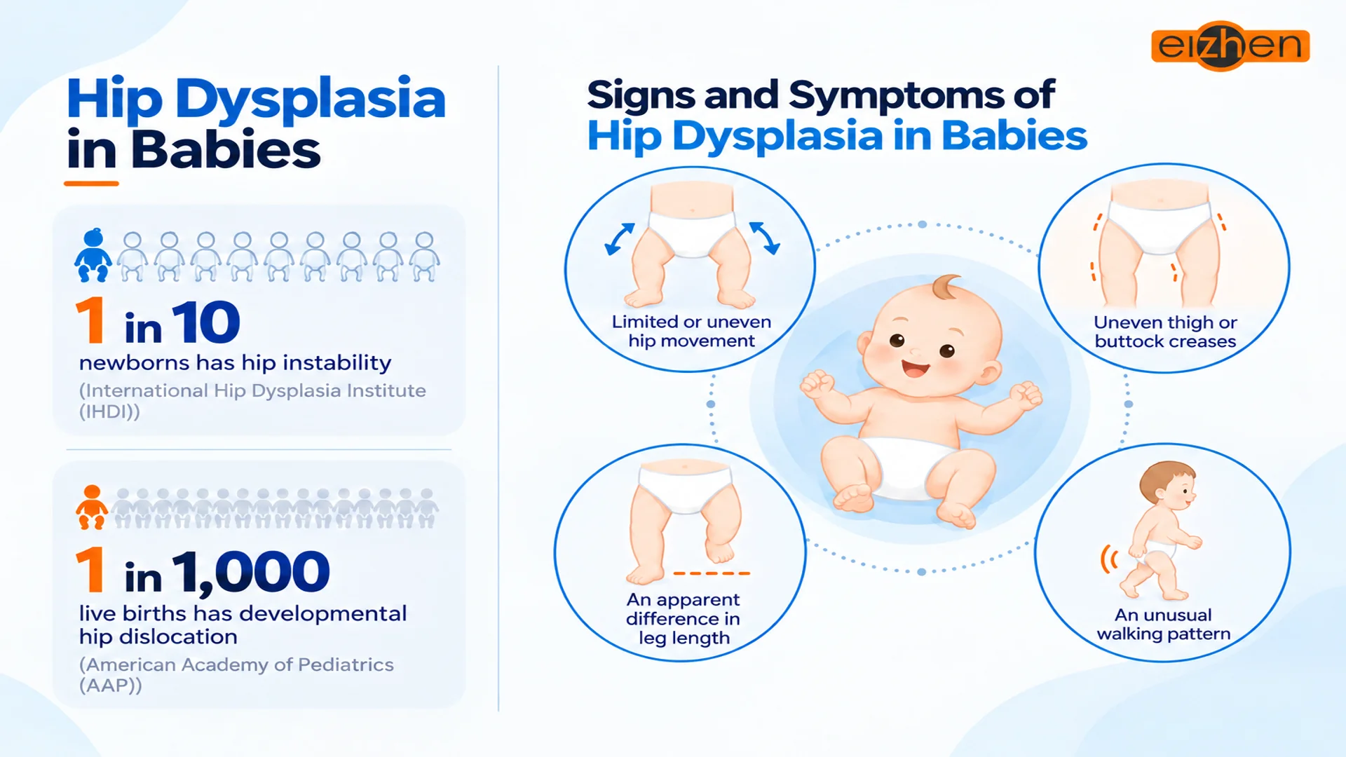
According to the International Hip Dysplasia Institute (IHDI), about 1 in 10 newborns has hip instability, and about 90% of these cases stabilize naturally after birth. However, early hip instability is not the same as the full spectrum of developmental dysplasia of the hip (DDH).
The American Academy of Pediatrics (AAP) estimates that developmental hip dislocation occurs in approximately 1 in 1,000 live births. This figure refers to hip dislocation rather than all forms of DDH, which can range from mild abnormalities to complete dislocation.
So, what signs may indicate hip dysplasia in babies?
DDH does not always show visible signs, so a baby may appear comfortable and move normally despite the condition. When signs are present, they may include:
- Limited or uneven hip movement: One hip may not move outward as freely as the other, particularly as the baby gets older.
- Uneven thigh or buttock creases: Skin folds may look different on each side, although this finding alone does not confirm DDH.
- An apparent difference in leg length: One leg may appear shorter than the other, particularly when the hips and knees are bent.
- An unusual walking pattern: Children with DDH who have started walking may develop a limp or a waddling gait.
What Causes Hip Dysplasia in Babies?

The exact cause of hip dysplasia in babies is not always clear, but several factors can increase a baby’s risk.
Family History and Genetics
Family history is an important risk factor. According to the International Hip Dysplasia Institute (IHDI), a child’s risk is approximately 12% if a parent has DDH. If both a parent and a previous child have the condition, the risk for another child rises to around 36%.
Factors During Pregnancy
A baby’s position during pregnancy can influence hip development and may cause abnormalities. Breech presentation, when the baby’s bottom or feet are positioned to emerge first, is a recognized risk factor, particularly late in pregnancy.
Reduced amniotic fluid may also contribute, although its association with nonsyndromic DDH is less firmly established. However, having these risk factors does not mean a baby will necessarily develop DDH.
Infant Positioning After Birth
Infant positioning after birth also matters. Tight swaddling that holds a baby’s legs straight and close together can restrict natural hip movement and increase the risk of DDH.
Although these factors can help explain a baby’s risk, DDH can occur without identifiable risk factors, making routine hip examinations important.
How Is Hip Dysplasia in Babies Diagnosed?
So far, we’ve explored what baby hip dysplasia is, its possible signs, and the factors that may increase the risk. But since DDH can be present without obvious signs, how do healthcare professionals identify it?
Diagnosis typically involves a physical examination and medical imaging tests, such as ultrasound scans or X-rays, depending on the baby’s age and clinical findings.
The main methods include:
- Physical examination: A healthcare professional checks the hips for instability, restricted movement, differences in leg length, and other findings that may suggest DDH.
- Infant hip ultrasound: This imaging method uses ultrasound machines and transducers to examine the hip joint and is particularly useful in young babies, whose hips contain substantial cartilage.
- X-ray: As the baby’s hip bones develop, a pelvic X-ray can help assess the position of the thighbone and the development of the hip socket; it is generally more useful from around 4–6 months of age.
| Diagnostic method | What it involves | When it is used | |
| Physical examination | Checks the hips for instability, restricted movement, leg length differences, and other signs that may suggest developmental dysplasia of the hip (DDH). | During routine infant examinations and when DDH is suspected. | |
| Infant hip ultrasound | Uses ultrasound waves to examine the hip joint, including the cartilage and the developing hip socket. |
|
|
| X-Ray | A pelvic X-ray helps assess the position of the thighbone and the development of the hip socket as the bones mature. | Becomes more useful around 4–6 months of age, although the choice between ultrasound and X-ray depends on the baby’s age and clinical findings. |
Among these methods, ultrasound machines and various types of transducers play an important role in evaluating young babies’ hips when imaging is indicated, allowing clinicians to assess structures not yet fully visible on X-rays.
In the next section, we’ll explore how infant hip ultrasound works and how it helps detect developmental dysplasia in babies.
Pediatric Hip Ultrasound and Probes: How They Help Detect Hip Dysplasia in Babies
As discussed in the previous section, pediatric hip ultrasound is one of the main imaging methods used to assess developmental dysplasia of the hip (DDH) in young babies.
During the examination, an ultrasound system and a compatible transducer produce real-time images of both hip joints, allowing clinicians to examine the cartilaginous femoral head, the acetabulum (hip socket), the acetabular roof, and the surrounding labrum. These views help assess hip socket development, femoral head positioning, and joint stability.
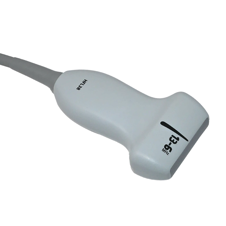
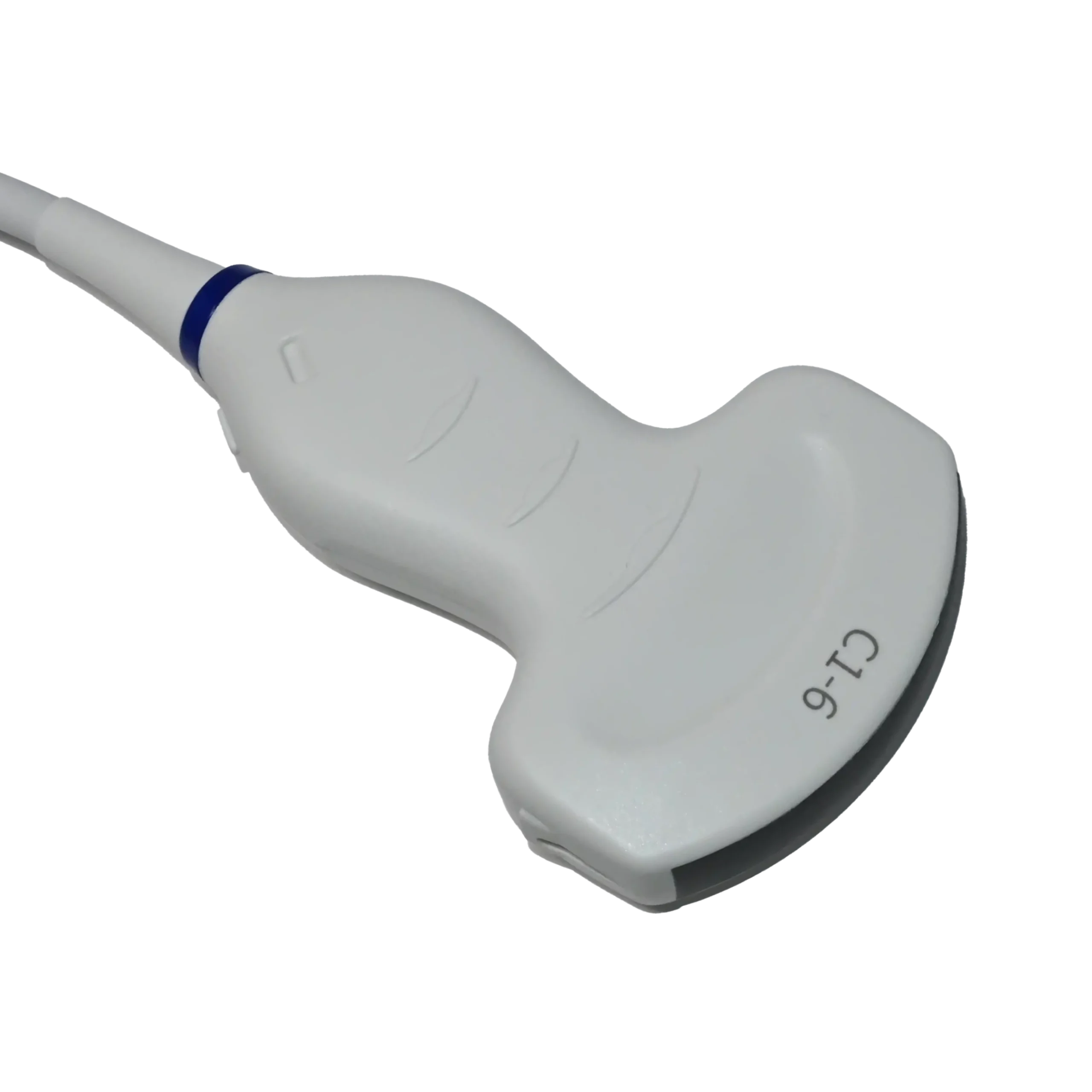
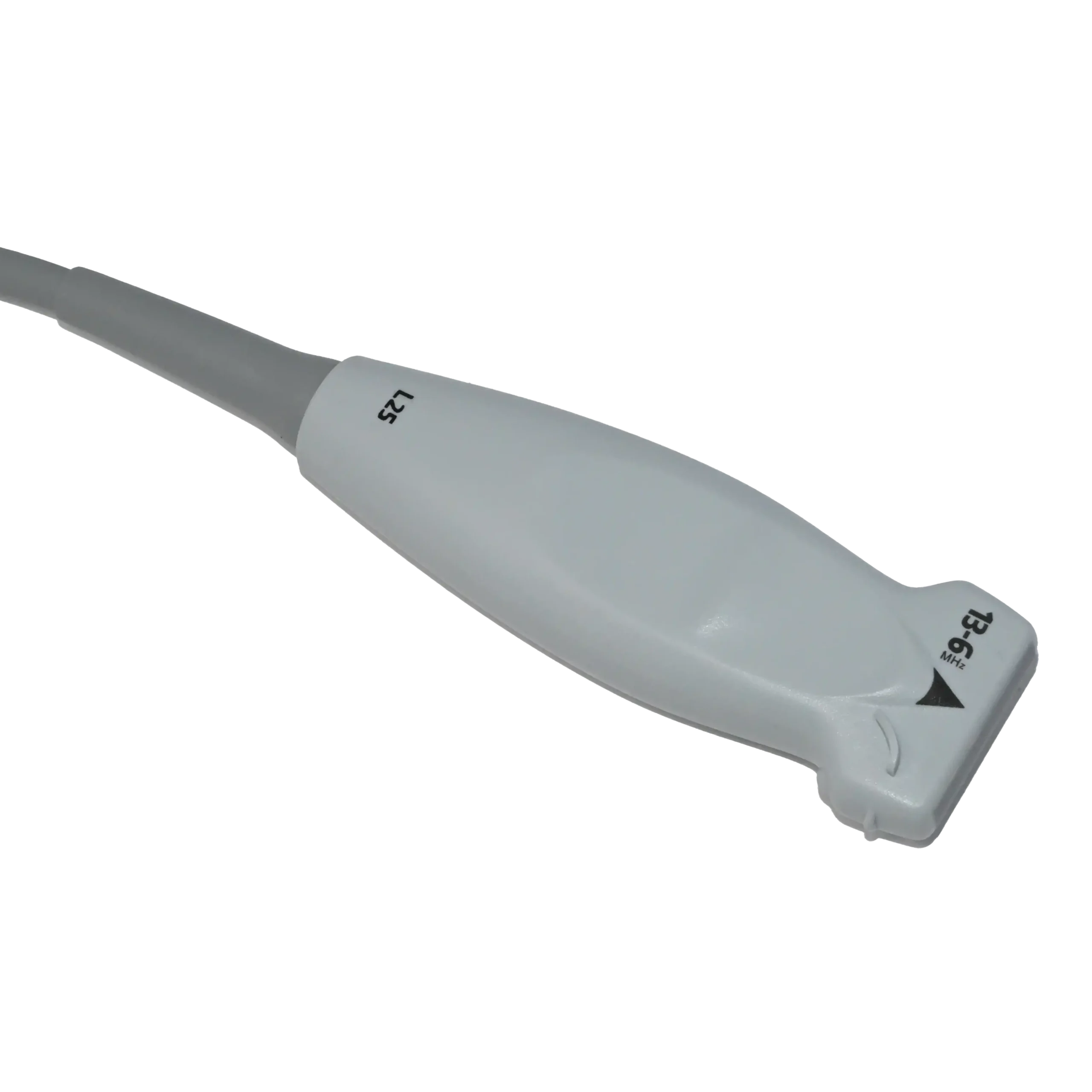
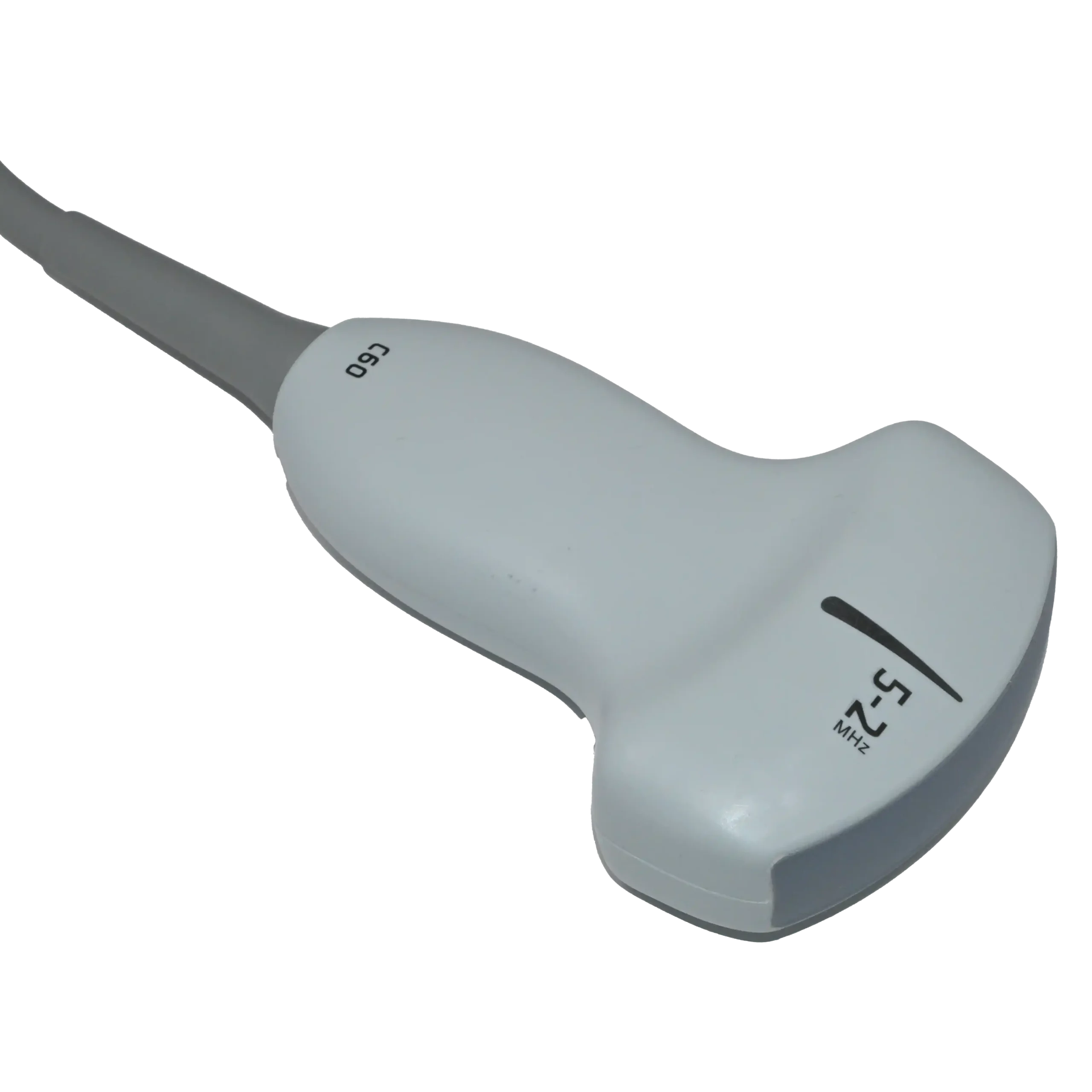
Ultrasound examinations of an infant’s hips require a transducer suited to the baby’s size and the structures being assessed. The most appropriate probe for detecting developmental dysplasia of the hip (DDH) is generally a high-frequency linear-array transducer, while other designs may be considered in selected circumstances, as explained below.
High-frequency linear-array transducer
According to the AIUM’s 2024 practice parameter, a high-frequency linear transducer is the primary choice for infant and pediatric hip ultrasound because it provides the detailed resolution needed to assess the femoral head, acetabulum, acetabular roof, and surrounding cartilage.
Curvilinear (convex) transducer
Although the high-frequency linear transducer is the standard probe for baby and pediatric hip ultrasound, the Radiology Department of Maastricht University Medical Center and Alrijne Hospital in the Netherlands note that a convex probe can also be practical in certain cases. Its curved footprint and broader field of view may help with certain patients.
What to Expect at a Pediatric Hip Ultrasound Test?
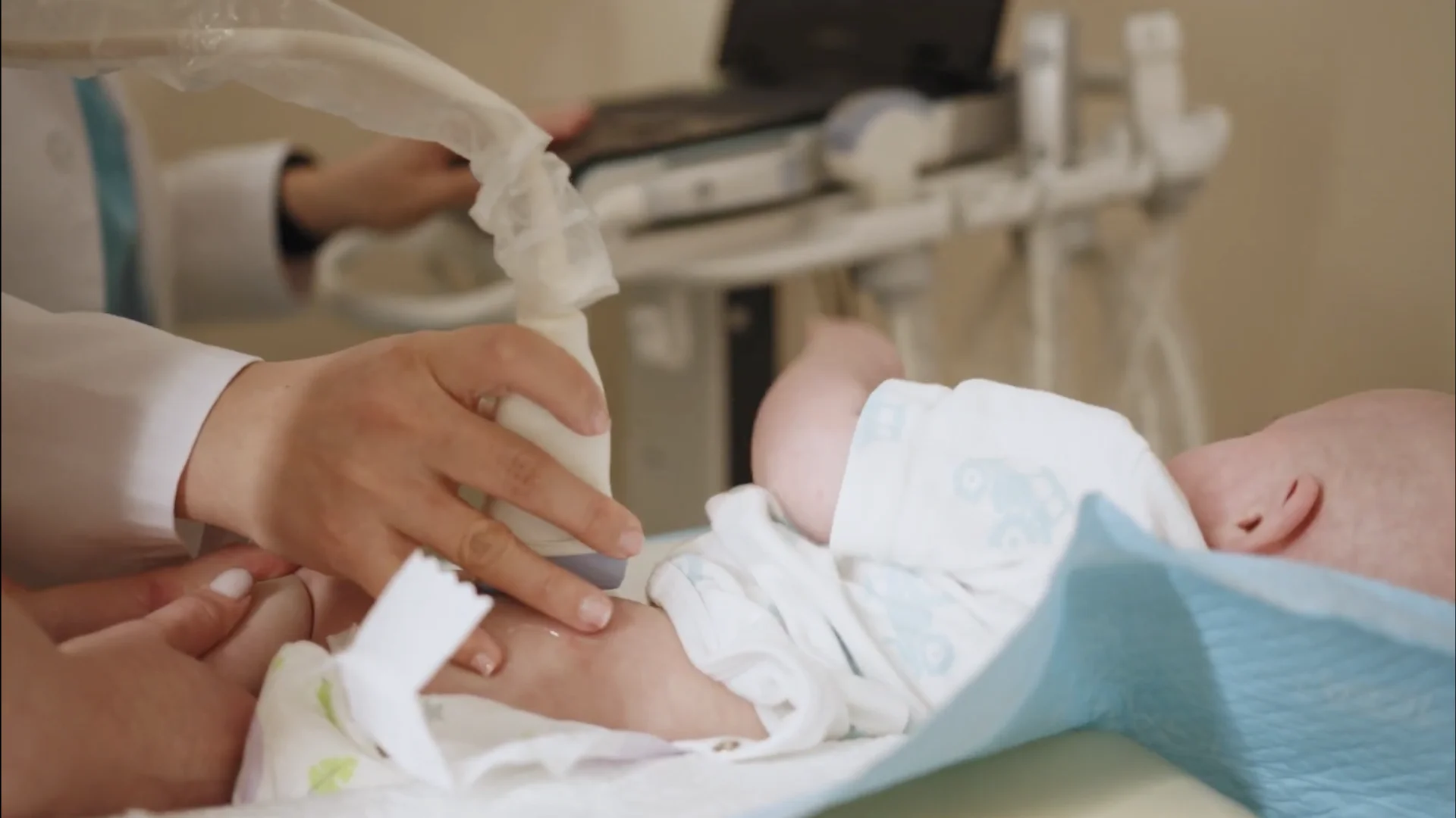
Before a pediatric hip ultrasound, a healthcare professional may ask about your baby’s medical history. To prepare for the scan, dress your baby in easily removable clothing, as the sonographer will usually need to uncover the lower body.
As shown in the image above, during the examination, your baby will lie on their side while the sonographer applies warm gel and gently moves the ultrasound transducer, commonly a linear probe, over both hips to capture images of the hip joints.
Because the scan is painless and uses no radiation, it is generally comfortable for babies. To help your baby stay calm, you can feed them beforehand. After the examination, the sonographer may discuss the initial findings with you or send the results to the referring clinician for further review.
Treatment Options for Hip Dysplasia in Babies
After DDH is identified through a physical examination and, when needed, imaging such as pediatric hip ultrasound, the next step depends on the baby’s individual condition.
Not every baby requires immediate treatment: mild, stable hip abnormalities may improve naturally and can sometimes be managed through regular examinations and follow-up imaging.
When treatment is necessary, the main goals are to guide the femoral head into the hip socket and support normal joint development. The most suitable approach depends on the baby’s age, hip stability, and the degree of displacement.
- Soft harness or brace: A Pavlik harness holds the baby’s hips in a position that helps keep the femoral head in the socket as the joint develops.
- Closed reduction and spica cast: A doctor guides the femoral head back into the hip socket without an incision, then holds it in place with a cast to maintain alignment.
- Surgery for more complex cases: When other treatments fail or aren’t suitable, surgery may be needed to reposition the femoral head or correct abnormalities in the developing hip bones.
| Age group | Medical Treatment | Other considerations |
| Newborns | Pelvic Harness | Follow-up examinations and imaging help assess whether the hip is responding to treatment. |
| 1–6 months (infants) | Pelvic Harness | A fixed abduction brace or another approach may be considered depending on hip stability and response to treatment. |
| 6–18 months | Closed Reduction or Spica Cast | If closed reduction is unsuitable or unsuccessful, open reduction using a medial or anterior surgical approach may be considered. |
Conclusion
In this article, we’ve explored hip dysplasia in babies, its possible signs and causes, diagnostic methods, and treatment options. Developmental dysplasia of the hip (DDH) occurs when the hip joint does not develop properly, potentially leaving the hip socket too shallow or the joint unstable.
When further imaging is needed, an infant or pediatric hip ultrasound is a valuable, radiation-free method for examining a baby’s developing hip joint using an appropriate transducer.
Early assessment helps healthcare professionals identify abnormalities and determine whether monitoring or treatment is needed. Depending on the baby’s age and hip condition, treatment may involve a Pavlik harness, closed reduction with a spica cast, or surgery in more complex cases.