An Overview of Different Types of Transducers
Ultrasound transducers are used to produce detailed images of the body’s internal structures. Different parts of the body require specific technologies, which is why there are various types of probes. Each type has its unique characteristics that allow doctors, clinicians, and healthcare providers to examine different areas of the body effectively. In this article, we take a closer look at the different types of transducers and their key features.
Key Features of Different Types of Transducers
There are different types of ultrasound transducers, each distinguished by unique characteristics. The key differences, including piezoelectric crystal arrangement, size, shape, frequency, and footprint, are explained below.
1- Piezoelectric Crystal Arrangement
Piezoelectric crystals are the essential components of ultrasound probes (or transducers) that generate and receive ultrasound waves. The arrangement of these crystals within the probe is crucial for its functionality and the quality of the ultrasound images produced.
2- Size and Shape
A key factor distinguishing different ultrasound transducer types is their shape and size. Transducers can vary significantly in design; some feature a flat, rectangular shape, while others are designed with a curved or convex shape. These shape variations are tailored to specific applications and imaging needs, providing advantages in how they fit the body’s contours or the type of image they produce.
3- Frequency
One criterion for comparing different types of transducers is their frequency. Ultrasound transducers come in both low- and high-frequency options, with frequency referring to the sound waves emitted by the probe. Higher frequencies generally offer superior image quality but do not penetrate as deeply as lower frequencies.
4- Footprint
Another important factor differentiating types of ultrasound probes (or ultrasound transducers) is their footprint. The “footprint” refers to the size and shape of the area on the probe that comes into contact with the patient’s body during an ultrasound examination. In other words, the physical surface of the ultrasound transducer directly touches the skin or the area being scanned. Some ultrasound probes are designed with small footprints for imaging in tight or hard-to-reach areas, while others have larger footprints to capture a broader area in a single scan.
What Are the Different Types of Transducers?
As mentioned earlier, there are now various types of probes, also known as transducers. From the early days of medical imaging, when only limited equipment was available, this field has advanced rapidly, offering a wide range of options today. Here, we explore the seven main types of transducers commonly used by clinicians and healthcare providers.
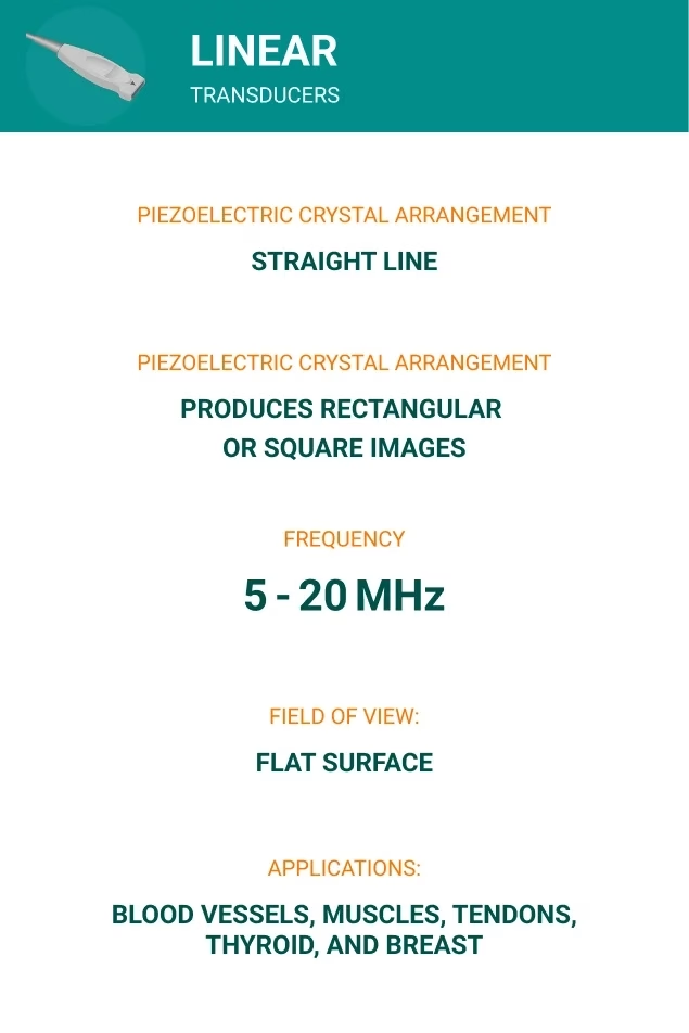
1- Linear Transducers
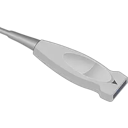
One type of ultrasound transducer is the linear transducer, which features a flat, rectangular shape.
- Piezoelectric Crystal Arrangement: These probes are equipped with piezoelectric elements arranged in a straight line to generate and receive ultrasound waves.
- Image Shape: The linear arrangement produces a rectangular or square image.
- Frequency: Linear transducers operate at high frequencies, typically ranging from 5 MHz to 20 MHz, which translates to high resolution and allows for detailed images of superficial structures.
- Field of View: Because linear transducers have a flat surface that produces a rectangular field of view, they can achieve consistent depth across the entire image.
- Applications: These types of ultrasound probes are ideal for imaging superficial structures like blood vessels, muscles, tendons, thyroid, and breast. They are commonly used in vascular, musculoskeletal, and small-part ultrasound.
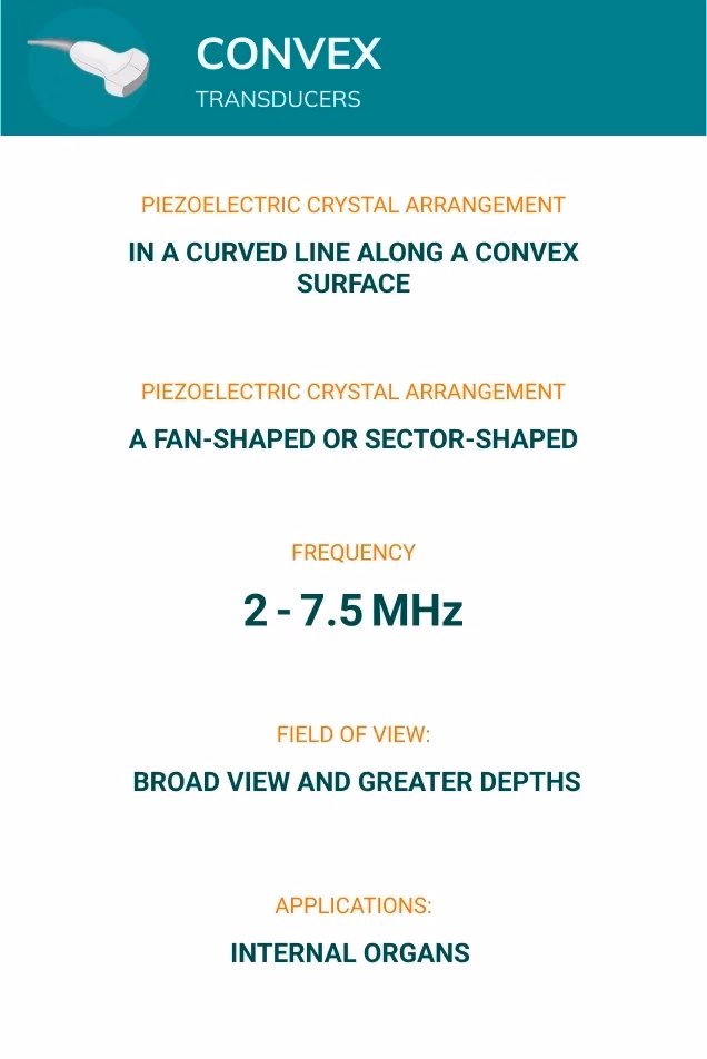
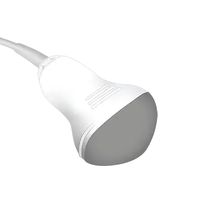
2- Convex Transducers (Curved or Curvilinear Probe)
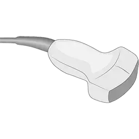
Another type of ultrasound transducer is the convex transducer, also known as a curved or curvilinear probe.
- Piezoelectric Crystal Arrangement: In convex probes, the crystals are arranged in a curved line, typically along a convex surface.
- Image Shape: They are designed with curved surfaces, creating a fan-shaped or sector-shaped field of view.
- Frequency: Image quality is lower than linear transducers, operating between 2 and 7.5 MHz, allowing for deeper penetration.
- Field of View: They provide a broad view with greater depth and moderate resolution.
- Applications: Convex probes are ideal for abdominal scans, providing a comprehensive view of internal organs.
3- Phased Array Transducers
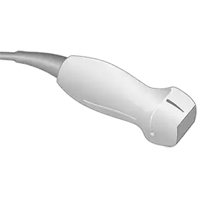
A phased array ultrasound transducer characterized by its small footprint and advanced technology.
- Piezoelectric Crystal Arrangement: Phased array probes contain 68 to 128 small crystals arranged in a linear or curvilinear pattern.
- Image Shape: They provide a pie-shaped (sector) image and compact design for imaging in tight or specific areas.
- Frequency: These ultrasound probes operate at low frequencies, typically between 2 MHz and 7.5 MHz.
- Field of View: They electronically focus and steer the beam for high-quality imaging.
- Applications: They are commonly used for cardiac examinations, including transesophageal echocardiography (TEE), and abdominal and brain examinations.
4- Pencil Transducers
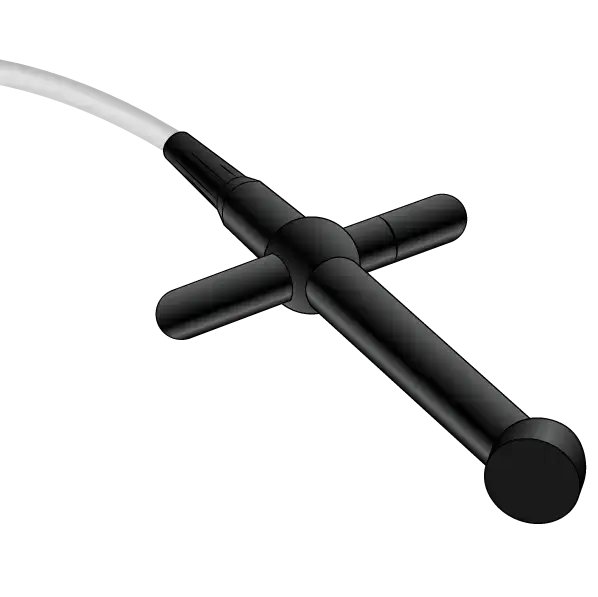
Pencil transducers, also known as pencil probes or CW (Continuous Wave), are a specialized type of ultrasound transducer commonly used in Doppler ultrasound studies, particularly for vascular and cardiac applications.
- Piezoelectric Crystal Arrangement: Unlike other types of ultrasound probes, which use a large array of crystals to create images, pencil transducers are equipped with only two separate piezoelectric crystals.
- Image Shape: As their name suggests, pencil transducers resemble the shape of a pencil with a small footprint.
- Frequency: They operate at low frequencies, typically ranging from 2 MHz to 8 MHz.
- Field of View: They are designed with a very narrow and focused field of view, aiming to target specific areas, such as a blood vessel or heart valve.
- Applications: These ultrasound probes are widely used in vascular studies to assess blood flow.
5- Endocavitary Transducers
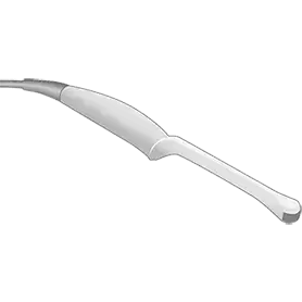
Endocavitary transducers are designed for internal imaging and fit into certain body orifices.
- Piezoelectric Crystal Arrangement: The piezoelectric crystals in endocavitary transducers are typically arranged in a curved linear array along the probe’s surface.
- Image Shape: They have a small footprint and a slender, elongated shape that makes them easy to insert into body cavities.
- Frequency: Endocavitary transducers operate at frequencies ranging from 3.5 MHz to 11.5 MHz
- Field of View: The curved arrangement of the crystals allows for imaging sector-shaped pictures to cover a wide area from a relatively small insertion point.
- Applications: They are widely used in transvaginal and transrectal exams and commonly employed in procedures like prostate biopsies and other ultrasound-guided internal exams.
6- Transesophageal TEE Transducers
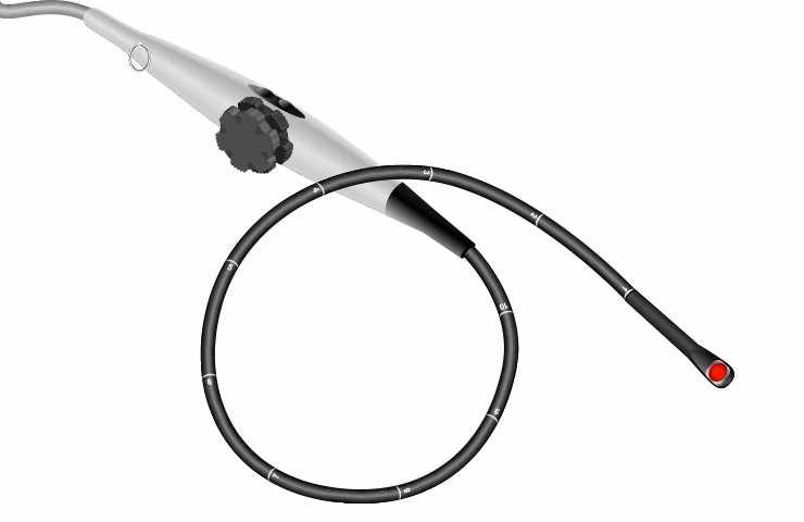
Unlike standard transthoracic echocardiography, which places the ultrasound probe on the chest, transesophageal echocardiography (TEE) involves guiding a flexible probe with a transducer down the esophagus. Since the esophagus runs directly behind the heart and major blood vessels, this technique allows for sharper, more detailed imaging.
- Piezoelectric Crystal Arrangement: In TEE transducers, the piezoelectric crystals are typically arranged in a phased array configuration.
- Image Shape: TEE transducers are formed with a Sector-shaped (fan-shaped).
- Frequency: TEE probes operate at mid-range frequencies, typically between 3 and 7 MHz.
- Field of View: These probes, which are sector-shaped, allow for the visualization of a wide area of the heart from the esophagus.
- Applications: TEE ultrasound probes are suitable for detailed cardiac imaging, intraoperative monitoring, and the assessment of complex cardiovascular conditions.
7- 3D/4D Transducers
So far, we’ve explored 2D ultrasound transducers, the most common types of ultrasound probes used in medical imaging, which produce two-dimensional images. However, advancements in technology have led to the development of 3D and 4D ultrasound probes. They offer more detailed and dynamic imaging capabilities compared to traditional 2D transducers.
- 3D transducers can provide three-dimensional images of structures within the body, making them particularly useful in prenatal imaging. They allow for greater precision in the detection of fetal abnormalities.
- 4D transducers enhance 3D imaging by adding the dimension of time, enabling the creation of “live” or real-time 3D images that visualize motion, such as a beating heart or the movements of a fetus in the womb.
The Importance of Choosing the Right Ultrasound
In the vast and varied field of medical imaging, different types of transducers come in various shapes, sizes, and with different diagnostic purposes. The selection of an appropriate transducer is largely determined by the area of the body that needs to be examined.
For instance, transducers with a broad view are ideal for examining deeper structures within the body. These probes are often used in abdominal scans or during pregnancy to visualize the fetus. On the other hand, small, high-frequency transducers are better suited for imaging superficial areas, such as small organs, blood vessels, or musculoskeletal structures. The more knowledge you have about these options, the better equipped you will be to make an informed decision. Understanding each transducer type’s unique features and advantages can greatly enhance diagnostic accuracy and patient care.
Beyond Transducers with Elzhen: Reliable Probe Solutions
Beyond just understanding the different types of transducers, ensuring their reliability is what matters most.
Whether you’re a clinician, doctor, or healthcare professional, having dependable ultrasound probes is essential for accurate diagnostics. At Elzhen, we provide refurbished probes that deliver reliable performance and excellent image quality. Every probe undergoes thorough technical testing, and you can conveniently order them online at Elzhen.com.
Already own a probe but need maintenance instead of a replacement? Our probe repair service is the perfect solution, helping you extend the life of your equipment without the cost of buying new.

Medical Imaging