Beyond the Baby Bump: The Hidden Power of Abdominal Ultrasound
When most people think of abdominal ultrasound imaging, the first thing that often comes to mind is pregnancy; a mother lying on a table, with a doctor using a specialized abdominal ultrasound device to check on the baby. While this is one of the most common uses of ultrasound technology, the role of these machines goes far beyond that.
Abdominal imaging is a powerful tool for examining a wide range of internal organs and conditions. In this article, we’ll explore the capabilities of abdominal ultrasound machines, dive into the world of abdominal imaging, and uncover how these scans can reveal hidden health secrets.
What is an Abdominal Ultrasound?
In our previous blogs, we discussed ultrasound machines, smart and innovative devices that truly transformed the medical imaging industry. These systems provide real-time visualization of different body parts while preserving patient safety at the highest levels.
We’ve also explored the different types of ultrasound machines, from portable models used in emergency rooms to console-based systems designed for advanced imaging.
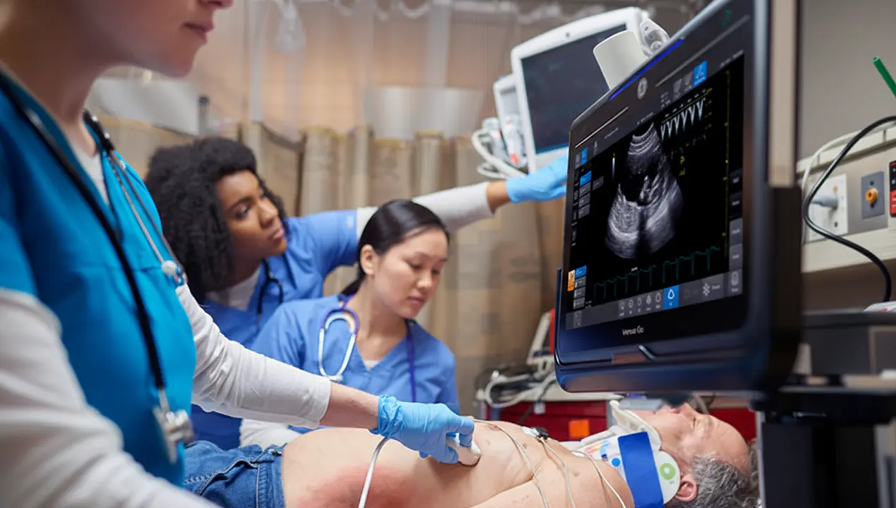
Now, let’s take a closer look at one of the most common and essential applications: abdominal ultrasound. Abdominal ultrasound is a non-invasive diagnostic technique to examine the organs and structures inside the abdomen.
When it comes to the equipment, transducers (or probes) play a key role in how clear and useful the images are. For abdominal ultrasound, the most commonly used transducers include:
- Curvilinear (or convex) probes: These are the most popular for abdominal scans due to their wider field of view and low-frequency range (typically 2–5 MHz). Convex or curvilinear transducers allow for deeper penetration and are ideal for visualizing large organs.
- Phased array probes: Phased array probes are primarily used for cardiac imaging. However, they also work well for abdominal scans in tight spaces, like when scanning through narrow windows.
- Linear probes: While not the first choice for full abdominal scans, linear transducers can be used to examine superficial structures.
How Does an Abdominal Ultrasound Work?
It’s essential to understand that ultrasound machines don’t directly produce images; this is where transducers play a crucial role.
The ultrasound machine generates and processes high-frequency sound waves, but it’s the transducer that acts as both the sender and receiver of those waves. Then, the machine takes those echoes and converts them into real-time images.
In simple terms, the machine provides the power and the brain, while the transducer acts as the eyes and ears! As a result, together, they create a dynamic imaging system that allows healthcare providers to safely and accurately view internal organs. Specifically for the abdomen, without requiring invasive procedures or radiation.
Meet the Machine: The Design of Abdominal Ultrasound Equipment
Abdominal ultrasound machines are available in two main forms: console-based systems and portable devices. Console-based machines are the larger, more traditional systems often found in hospitals and imaging centers. Some top models of console-based machines for abdominal imaging are GE HealthCare LOGIQ E10, Philips EPIQ 7, and Siemens Acuson S3000.
As below, they have a tall, rectangular structure, resembling a slim cabinet on wheels.
On the other hand, portable machines feature a lightweight and ergonomic design. Thanks to their physical shapes, portable machines are commonly used in emergency rooms, small clinics, ambulances, and even in-home care settings. The top models of portable devices for abdominal imaging are GE Healthcare Vscan and Philips Lumify. Some images of portable devices are below.
What Parts of the Body Does Abdominal Ultrasound Diagnose?
As its name suggests, an abdominal ultrasound machine focuses on imaging the abdominal area, the central region of the body that houses many vital organs. Therefore, it allows healthcare providers to examine the structure, size, and overall condition of the following abdominal organs:
- Liver: to detect fatty liver, tumors, cysts, or signs of hepatitis.
- Gallbladder: commonly checked for gallstones or inflammation (cholecystitis).
- Pancreas: to observe inflammation (pancreatitis), masses, or structural changes.
- Kidneys: for evaluating kidney stones, cysts, or signs of obstruction or infection.
- Spleen: to examine size changes, injuries, or underlying conditions.
- Bladder: to detect issues like urinary retention, tumors, or bladder wall thickening.
- Abdominal aorta: check for aneurysms or abnormal enlargement.
When and Why Doctors Order Abdominal Ultrasounds?
Doctors use an abdominal ultrasound to examine internal organs when they suspect something might be wrong with the abdomen. This is similar to when they use a cardiac ultrasound (echocardiogram) with TEE probes to examine the heart when a heart problem is suspected. They use specialized ultrasound machines designed for different medical applications in both cases.
Here are some of the most common reasons doctors use an abdominal ultrasound:
- Swelling or bloating: To check for fluid accumulation or enlarged organs.
- Abnormal blood test results: Particularly irregular liver or kidney function tests.
- Suspected gallstones or kidney stones: To confirm their presence and determine their size.
- Follow-up on previous findings: To assess changes in a known mass or track treatment progress.
- Urinary problems: To evaluate bladder function and identify potential obstructions.
- Aneurysm screening: Especially important for older adults at risk of abdominal aortic aneurysm.
How is the Abdominal Ultrasound Preparation
So far, we’ve discussed several topics, such as what an abdominal ultrasound is and how it works. But how would you be prepared as a doctor or a patient?
As a patient, you need to follow the tips below:
- Fast for 6 to 8 hours before the exam: In some cases, fasting reduces the gas in your digestive system and helps the doctor examine you with clearer images. However, in other cases, for example, for a routine pregnancy ultrasound, fasting is usually not required.
- Follow any special instructions: Your doctor might give you a specific diet the night before. Try to stick to it carefully, especially if you have a liver or gallbladder examination.
Continue medications if allowed: If you have taken any medicine, you might continue taking it. However, always check with your doctor first. - Wear loose, comfortable clothes. Depending on the area being scanned, you might need to change into a gown.
As a doctor or medical team, you should:
- Review the patient’s history and referral notes: This helps you understand the reason for the ultrasound examination and ensures you focus on the relevant organs.
- Prepare and check the equipment: Before starting, make sure the ultrasound machine is properly cleaned and disinfected. Also, remember to inspect the transducers.
- Communicate clearly: Maintain patient confidentiality and explain each step of the procedure. This helps the patient feel more informed and at ease.
Using Abdominal Ultrasound During Pregnancy
One of the main situations in which doctors use an abdominal ultrasound machine is when there is a baby! These machines help doctors check the baby’s heartbeat, growth, movement, and position while also examining the placenta, amniotic fluid, and uterus. Ultrasound imaging is a noninvasive method to ensure the pregnancy progresses normally.
Although pregnancy can bring joy and excitement, it also comes with potential health concerns. Problems like unexpected bleeding, severe cramping, lack of fetal movement, or concerns about the baby’s growth or position may arise. In these situations, doctors can rely on abdominal ultrasound and pregnancy probes as safe and effective tools to monitor both the mother and the baby.
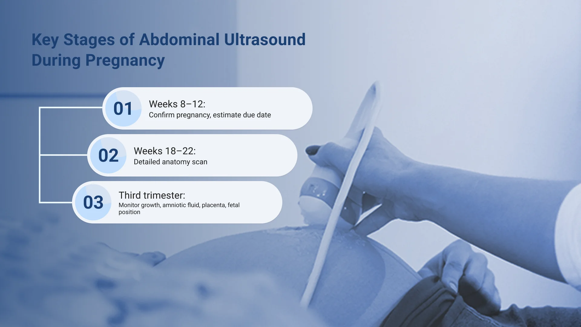
Doctors typically do an abdominal examination using ultrasounds at key stages:
- In the first trimester (around weeks 8–12): To confirm the pregnancy and estimate the due date.
- During weeks 18–22: To perform a detailed anatomy scan to check the baby’s organs and development.
- In the third trimester: To monitor fetal growth, position, or complications like low amniotic fluid or placenta previa.
What to Expect During an Abdominal Exam?
Doctors often recommend an abdominal ultrasound test to check for issues like unexplained pain or bloating, pregnancy monitoring, gallstones, kidney stones, or liver problems. Here’s what happens during the exam:
- Follow pre-exam instructions
As mentioned earlier, your doctor may ask you to fast for a few hours before the scan. This improves the quality of the images and helps get more accurate results. - Lie down on the exam table
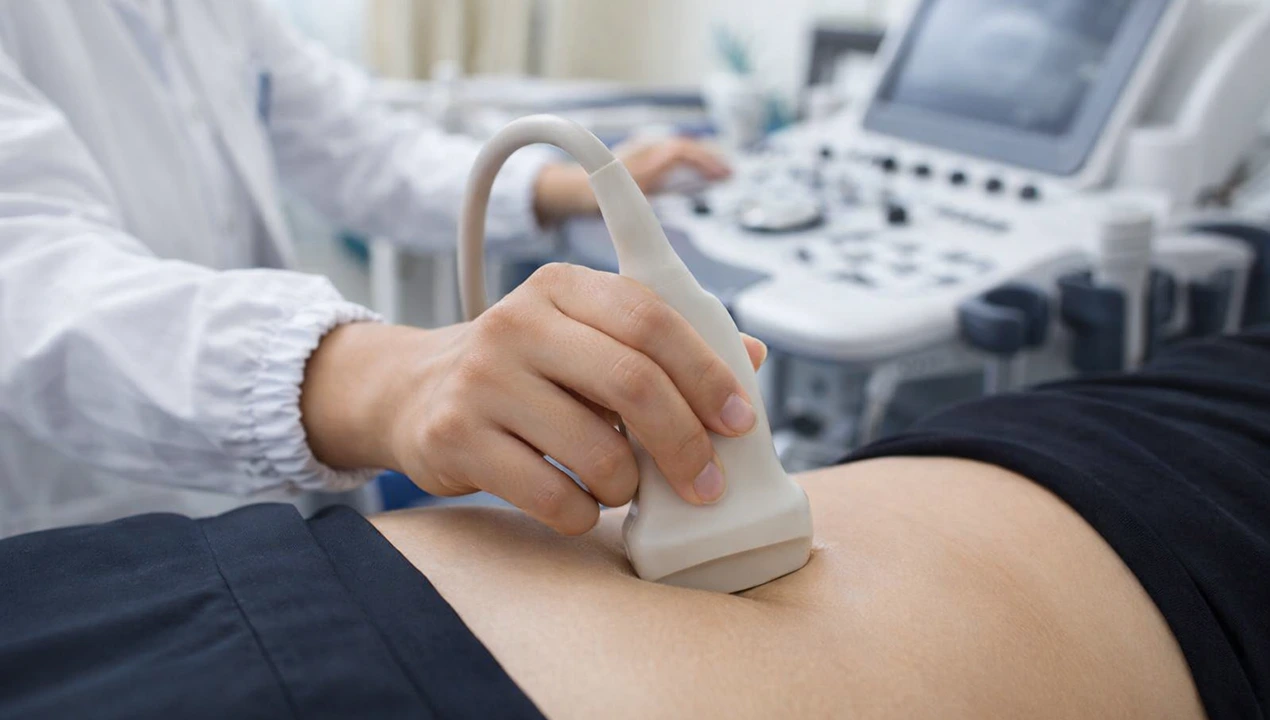
When you go for the exam, the technician will ask you to lie down in a specific position, depending on which part of your abdomen needs to be scanned. - Apply the ultrasound gel
Next, the technician uses a special gel on your abdomen. This gel helps the ultrasound probe move smoothly and allows it to capture clear images. - Move the probe over your skin
Then, the technician gently moves the probe across your abdominal skin. As they scan, you’ll see real-time images of your internal organs on a screen or ultrasound monitor. - Wipe off the gel and get ready to go
Once the technician finishes the exam, you can remove the gel, get dressed, and return to your normal activities immediately; no recovery time is needed. - Receive your results
Finally, a doctor or radiologist reviews the scans and sends the results to your healthcare provider, who will explain the results.
How Accurate is Abdominal Ultrasound Imaging?
Although ultrasound machines are the core devices, they are not enough by themselves, and additional equipment is necessary for comprehensive imaging. Several key factors must work together to ensure high-quality and accurate imaging, from skilled operators to proper machine settings and functional ultrasound transducers. When it comes to medical imaging, the transducer (probe) is what sends and receives the ultrasound waves, and it has a real impact on image quality and accuracy.
Imagine trying to perform a test with a damaged probe; the results would be unreliable. So, before using abdominal ultrasound machines or other types, it’s essential to ensure the transducers are working properly. Fortunately, you don’t always need a technician to assess the probe. In today’s world, breakthrough innovations like a smart transducer analyzer allow you to test your ultrasound probes quickly and reliably, without needing a PC or extra tools. This ensures better imaging and improved patient outcomes.
Conclusion
That’s all about abdominal ultrasound machines, their medical applications, and other relatable details. As mentioned, these machines are essential for examining various internal organs, including the liver, gallbladder, kidneys, and bladder. These devices’ most common ultrasound probes are convex or curved arrays. You can find more information about abdominal ultrasound machines and their compatible transducers at Elzhen.